Fig. 12.1
Trial posterior-stabilized components (Triathlon(®) (Stryker, Kalamazoo, MI, US)
The cam and post design of the PS TKA was introduced in 1978 to aid in knee flexion by facilitating posterior femoral rollback [1, 5, 6]. In addition to improving range of motion (ROM), the PS design was created to improve stair-climbing ability and prevent posterior subluxation of the tibia [6]. As mentioned above, the PS design employs a cam and post which when engaged during flexion prevents anterior translation of the femoral component on the tibia, causing femoral rollback, theoretically increasing flexion [1]. To evaluate the kinematics of PS designs, studies of native and TKA kinematics have been performed in a number of ways using cadaveric models, gait lab studies using cameras and skin markers, quasi-dynamic MRI, and computer-aided dynamic fluoroscopy. However, due to nonstandardized testing regimens, conflicting data have been reported regarding the cam-post interaction and femoral rollback in PS implants [4]. Variations in results can be explained by a number of uncontrolled factors limiting inter-study comparisons, including the positioning of the post in relation to the tibia. A posteriorly translated post (either by polyethylene design or surgical implantation) would cause earlier engagement and therefore theoretically increase posterior femoral translation and rollback in deep flexion. In comparison, an anteriorly translated tibial post would result in late engagement in flexion and potentially early disengagement of the cam and spine at high flexion angles [2, 4].
Li et al. performed an in vitro cadaveric examination of PS knees [4]. This group compared native knees before and after implantation of Zimmer NexGen© LPS TKA (Zimmer, Warsaw, IN, USA) using a robotic system to apply standardized loads to different muscle groups in various degrees of flexion from 0° to 120°. The study showed significantly decreased posterior femoral rollback of both the lateral and medial femoral condyles with the PS TKA compared to the native knee, achieving only 80 % of the native knee femoral rollback once the tibial post engaged the cam in deep flexion. This study also found decreased axial tibial rotation at 30° and 60° of flexion compared to native knee but similar tibial rotation at 90° and 120° of flexion [4].
Increased polyethylene wear and debris from the mechanical articulation of the cam and post is an area of concern with PS designs; in fact, a number of retrieval studies have shown the wear at the cam-post engagement interface to be significant [7, 8]. As a result, numerous component design changes have been made to help reduce the cam-post impingement, in addition to improvements in polyethylene thickness, sterilization, and manufacturing [7]. While implant design changes have been made, wear patterns on the post are highly dependent on proper ligamentous balancing and component alignment at the time of implantation [7, 8]. For example, anterior impingement of the post and box can also occur with knee hyperextension with a loose extension gap [8]. More constrained PS implants often have higher medial and lateral post wear due to the nature of the more constrained implant (often a result of a tighter articulation in the box between the femoral condyles and post) and lack of ligamentous stability for which that type of implant was chosen [7].
In a study by Dennis et al., video fluoroscopy was used to examine in vivo native, PS, and CR knees. In this study, average non-weight-bearing flexion values were similar between the PS and CR groups at 127° and 123°, respectively, but less than the native knees at 139° [9]. However, with weight-bearing deep knee bend, PS TKA achieved significantly higher flexion than CR knees (normal 135, CR 103, and PS 113) despite PS patients having a trend toward less preoperative ROM and lower clinical performance ratings preoperatively [9]. They concluded that in the non-weight-bearing setting, the primary constraints to joint laxity and knee flexion are the surrounding soft tissues in contrast with weight bearing where the geometric conformity of the implant surfaces acts as a primary restraint to joint laxity [9].
12.3 Cruciate Retaining (Fig. 12.2)
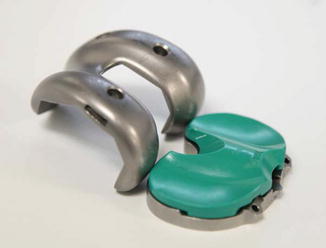
Fig. 12.2
Trial cruciate-retaining components (Triathlon(®) (Stryker, Kalamazoo, MI, US)
In an in vitro cadaveric study by Most et al., NexGen© LPS and CR TKA (Zimmer, Warsaw, IN, USA) implants were compared to native knee kinematics prior to implantation [2]. In this study, the CR knees achieved 80 % of native femoral rollback at 120° of flexion with an intact PCL. However, a significantly decreased posterior translation of the lateral femoral condyle and an increase in paradoxical anterior translation of the medial femoral condyle at flexion angles of 90° and 120° were observed when the PCL was resected. The PS TKA behaved similarly to the PCL-deficient CR TKA at 0–60° but then achieved 80 % of posterior femoral rollback at 120° of flexion, similar to the CR TKA. Thus, the authors concluded that the PCL contributes to posterior femoral translation in CR TKA at flexion angles greater than 30°. Internal tibial rotation was similar between PCL-retaining and PCL-deficient knees, signifying that the PCL plays more of a roll in posterior translation than in the screw home mechanism of internal tibial rotation that is more likely controlled by implant geometry and surrounding soft tissue constraints [2]. Contributing to the difference in kinematics between CR and PS knees, the CR femoral component has a larger radius of curvature on the medial side to help promote the internal rotation of the tibial component during flexion compared to the symmetric radii of curvature of the PS implant system [2]. A theoretical disadvantage of CR design is that the arthritic, poor-functioning, diseased PCL at the time of surgery may not function as intended to prevent anterior translation of the femur in flexion [10].
12.4 Medial Rotation (Fig. 12.3)
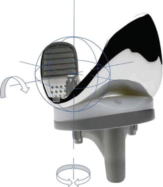
Fig. 12.3
The SAIPH® Knee (MatOrtho® Ltd., UK), an example of a medial ball and socket arthroplasty prosthesis
The MR design differs from the aforementioned CR and PS designs by a characteristically designed tibial polyethylene insert with high conformity to the medial femoral condyles [11]. The design consists of a ball-in-socket medial bearing and a less conformed lateral trough with the goal of creating a stable medial compartment and a mobile lateral compartment facilitating femoral rollback and internal tibial rotation [12, 13].
In a computer-modeled TKA study, an MR design was implanted in a normal, nonarthritic knee and was compared to PS, CR, or CR without an intact PCL knee to evaluate the necessity of a cam-post or an intact PCL for the medial pivot prosthesis [11]. They concluded that the high-conformity insert could not stand alone in attempting to restore normal kinematics including internal tibial rotation in a medial rotation fashion nor sufficient posterior femoral rollback [11].
In a randomized controlled trial by Hossain et al., an MR TKA was compared to a conventional PS implant and analyzed for post-op ROM and functional outcome scores. The Medial Rotation Knee© group had significantly higher ROM (98.2° vs. 115.5°) and improvements in the WOMAC pain subscale, SF-36 physical component, and TKFQ. They attributed the difference in ROM and outcomes to a variable engagement of the cam and post of the PS implant in comparison to the smooth relatively constrained medial compartment of the MR implant design. The authors concluded that the MR design better emulates the native knee modified hinge joint with a medially based rotational axis, when compared to the PS design, potentially translating to more efficient knee kinematics and the observed increased ROM [5]. A similar advantage to a CR design, an MR implant without a PS component eliminates the need to remove the native femoral bone for the box cut, thus preserving the bone. Further, because there is no cam-post mechanism, it is believed that this design may result in lower contact stresses and less polyethylene wear, since the majority of wear in PS designs occurs on the post [7, 14, 15].
12.5 Discussion
Clinical studies have not demonstrated significant differences in success or postoperative knee flexion angles between PS, MR, and CR TKA implant designs [2, 16]. While kinematic studies are useful, an important limitation of in vivo kinematic studies is the inability of obtaining preoperative kinematics of the intact native knee prior to implantation of TKA [2]. Still, more variable kinematics have been seen in CR compared PS designs [17], including “paradoxical” medial condylar anterior motion as well as a less consistent and lower-magnitude posterior femoral rollback of the lateral condyle [16, 18–20]. In a prospective randomized trial, Victor et al. compared PS and CR TKA kinematics and outcomes using a geometrically similar implant. PCL intact knees determined intraoperatively were then randomized to either a geometrically similar PS or CR Genesis II© implant [16]. No significant differences in clinical outcomes out to 5 years were found between the two groups. They found no significant differences in knee ROM in both passive and WB lunge activity testing. They demonstrated a statistically significant anterior medial slide in CR of 4 mm in comparison to posterior translation in PS of 3 mm and a greater lateral condylar posterior translation in the PS group. Both implant types demonstrated the “screw home mechanism” with internal tibial rotation during knee flexion. However, the PS design demonstrated an overall posterolateral translation consistent with a medial pivot axis, whereas the CR design had greater medial anterior translation, resulting in a central pivot axis [16]. The similar total amounts of axial internal rotation between the two implant designs are explained by the different motions of the two implants with the rotation created by a majority of posterolateral rotation of the lateral condyle in PS vs. a combination of medial anterior slide and posterolateral rotation of the lateral condyle in the CR knee [16].
Related posts:
 Aetiology of Patient Dissatisfaction Following Total Knee Arthroplasty
 Technological Aids in Total Knee Arthroplasty: Navigation, Patient-Specific Instrumentation, and Robotics
 Longevity: Characteristics of a Well-Functioning, Long-Lasting Total Knee Arthroplasty
 Primary Knee Arthroplasty: The Patella-Resurfacing Options
 Periprosthetic Fractures
 Revision Total Knee Arthroplasty: Surgical Technique in Dealing with Extensor Mechanism Failure
Stay updated, free articles. Join our Telegram channel

Full access? Get Clinical Tree


