Current efforts for prevention of hip dysplasia are primarily focused on early detection and early intervention to avoid long-term consequences of neglected hip dysplasia. True prevention efforts would eliminate the disorder before it develops. Better prevention may be possible by decreasing postnatal environmental factors that influence the development of hip dysplasia. This article reviews the natural history, prevalence, and etiology of hip dysplasia along with current methodologies for early diagnosis and possible considerations for prevention of neonatal hip instability and adult acetabular dysplasia.
- •
Current efforts for prevention of hip dysplasia are primarily focused on early detection and early intervention to avoid long-term consequences of neglected hip dysplasia.
- •
Better prevention may be possible by decreasing postnatal environmental factors that influence the development of hip dysplasia.
- •
Methods of prevention have focused on neonatal hip instability although 90% of adult acetabular dysplasia is unrecognized during childhood.
Early diagnosis and early treatment of congenital dislocation of the hip are now possible. The next great challenge is the prevention of this serious and disabling condition. —Robert B. Salter, Alexander Gibson Memorial Address, University of Manitoba, October 27, 1967
Introduction
A discussion of the prevention of developmental dysplasia of the hip (DDH) in children and adults must first define the term, disease prevention . This simple point is important because current approaches often focus on early diagnosis and treatment to prevent adverse long-term effects. In contrast, true disease prevention seeks to eradicate the disease so that the treating physicians never encounter the problem. An obvious example is the eradication of smallpox or elimination of polio in Western countries. This article considers opportunities for early detection in addition to possibilities for true prevention of hip dysplasia. A brief review of the natural history, prevalence, and etiology is used to frame the discussion of prevention.
The natural history of developmental dysplasia of the hip
Perhaps the best description of the long-term effects of untreated hip dysplasia is that of Canadian Indians observed in Manitoba by Corrigan and Segal in the 1940s:
The senior author first visited Island Lake in the summer of 1940 to attend as the government doctor at the annual treaty payments. He had never seen so many cripples all gathered together in one place outside of a hospital. It was easy to see that a large number of the cripples were cases of congenital dislocation of the hip, some bilateral, some unilateral. Some crawled on their hands and knees, some hopped about like clowns, others waddled like ducks. All accepted with typical stoicism their misfortune as life’s lot. They knew no different. Nature had exacted another toll.
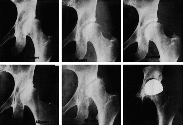
There are few scientific reports of the long-term natural history of untreated DDH. Wedge and Wasylenko provided some insight in a study of 54 adults with unilateral or bilateral dysplasia or frank dislocation. These investigators reported that a false acetabulum correlated with lower modified Harris hip scores because of degenerative changes of the femoral head and false acetabulum. Patients with dysplasia and subluxation had more disability and earlier onset of osteoarthritis than those with frank dislocations ( Fig. 1 ). Overall, the investigators reported that 60% of the hips had poor outcomes. This high rate of poor outcomes might be assumed to increase with longer-term follow-up because half the patients were younger than age fifty at time of follow-up. There is no longer any doubt that treatment improves the natural history of DDH, but untreated hip dislocations still occur in remote regions of countries, such as Mexico, Ecuador, and Iraq.
Disease prevalence, scope, and importance
Hip dysplasia affects approximately 1% to 3% of newborn infants although 4% to 6% may be affected if milder forms are included. The combined total of all heart anomalies exceeds the incidence of hip dysplasia, but the specific diagnosis of hip instability is the single most common abnormality in newborn infants. Approximately 2 per 1000 (0.2%) infants have frank hip dislocation that can be detected at the time of newborn examination. In addition, ultrasound studies have demonstrated dynamic hip instability and/or ultrasonographic hip dysplasia in 5% to 15% of all newborns. Approximately 80% of mild cases of hip instability resolve spontaneously, but evaluation or treatment is recommended for 2% to 3% of all children because of concerns regarding hip dysplasia. Based on the number of new births in the United States each year, it is estimated that approximately 100,000 infants in this country undergo additional evaluation for or treatment of hip dysplasia each year. Although ultrasound screening has been criticized as overly sensitive, it may provide some insight into cases of predisposition to adult hip problems because 90% of adult hip dysplasia is undetected by clinical screening. Thus, the true prevalence of infantile hip dysplasia is difficult to assess and depends in part on the definition of dysplasia and the population reported.
In the adult population, hip dysplasia is a substantial contributor to the development of hip arthritis. Although primary osteoarthritis has been previously been identified as the most common cause of hip arthritis, this concept has been challenged by studies reporting a high prevalence of pre-existing hip deformity, such as acetabular dysplasia, as well as the pistol-grip deformity associated with slipped capital femoral epiphysis or femoroacetabular impingement. Solomon performed a clinical and radiographic analysis combined with postmortem examination of 327 cases of osteoarthritis of the hip. He identified acetabular dysplasia in 20%, with a male:female ratio of 1:10. In a radiographic study of primary osteoarthritis, Harris estimated that 40% of cases were caused by underlying hip dysplasia. Harris also noted the preponderance of women with acetabular dysplasia. Hoaglund and Steinbach summarized several historical articles, concluding that hip dysplasia accounts for approximately 10% of osteoarthritis of the hip. Using Hoaglund’s more conservative estimate of 10% of cases of osteoarthritis attributable to hip dysplasia translates to approximately 25,000 total hip replacements (THRs) a year in the United States as a result of hip dysplasia.
Hip dysplasia plays an even greater role in the need for THRs in young people. Engsaeter, et al in Norway reported that hip dysplasia is the origin of arthritis, requiring THR for approximately 20% of people younger than 40 years of age, and that 87% of those patients are women. In this study, 92% of patients with dysplasia requiring THR were undetected during childhood even though a national program for screening had been implemented in 1967. The Norwegian registry data also indicate that patients with neonatal hip instability (NHI) had a 2.6-times increased risk of early THR compared with children without NHI, although after treatment of NHI, numbers yield a low overall risk for THR before age 40. Although the data from Norway are useful, it is difficult to estimate the prevalence of unrecognized hip dysplasia in North America. If certain assumptions are made, then approximately 350,000 adults in the United States older than 40 are at risk for early hip arthritis because of dysplasia. This estimate is based on the prevalence of osteoarthritis of the hip in Western countries combined with the estimated rate of dysplasia as a cause of osteoarthritis. By age 40, 1.3% of the population has osteoarthritis of the hip. This percentage rises to 14% after age 85 ( Fig. 2 ). The prevalence of hip arthritis for the entire adult population is 3% to 8%. When these numbers are applied to the 140 million adults older than 40 in the United States, and 10% of the hip arthritis is caused by dysplasia, then approximately 350,000 adults older than 40 are at risk for early hip arthritis because of dysplasia.
To represent the spectrum of hip dysplasia, it may be advisable to distinguish between NHI and acetabular dysplasia that becomes evident later in life. Perhaps the etiologies are different or perhaps the adult type of hip dysplasia was stable but undetectable during infancy. If prevention of NHI and adult acetabular dysplasia is possible, then the neonatal period is the logical time for preventive measures. To consider prevention, the etiology of hip dysplasia may provide an understanding of possible interventions to reduce the burden of disease for this common condition.
Etiology of hip dysplasia
The role of inheritance in DDH is well known but hormonal and mechanical factors also affect the risk of NHI. Inheritance may predispose infants to the adverse influence of hormonal or mechanical factors. Thus, the etiology is multifactorial but there is a 12-fold increase in risk for first-degree relatives. If one child has hip dysplasia, the risk increases to 6% for a second child. The child of a parent with hip dysplasia has a 12% risk of having dysplasia. The risk is 36% for subsequent children when there is an affected parent and an affected child. DDH is 4 to 5 times more prevalent in girls than in boys. Girls may be especially susceptible to the maternal hormones estrogen and relaxin, which contribute to ligamentous laxity with resultant instability of the hip in the neonatal period.
There are also risk factors related to intrauterine mechanical pressure, including breech position, oligohydramnios, increased birthweight, prim parity, older maternal age, and postmaturity. Leutekort and colleagues reported that hip joint instability was present in 47% of breech position babies born vaginally with the knees extended, whereas hip instability was noted in only 8% of vaginally delivered breech infants whose knees were flexed. The protective effect of knee flexion, younger gestational age, and lower birthweight has also been observed in twin births. Breech infants born by caesarian section have a lower incidence of DDH than breech infants born vaginally. These findings suggest that stretching of the hip capsule or hamstring muscles in utero predispose to hip dislocation in the neonatal period.
Postnatal mechanical factors also affect the development of hip dislocation. Hip dislocation has been produced in experimental animals by immobilization of the hips or knees in extension, but sectioning of the hamstring or psoas muscles reduced the frequency of hip dislocation. In the experimental using animal models, the incidence of hip dislocation was also increased by addition of maternal hormones that promote hip joint laxity, and the effect was greater in female than in male animals.
Normal human infants have an average hip flexion contracture of 28°. The hip flexion contracture decreases to 19° at 6 weeks and 7° at 3 months of age. Hip flexion contractures of 50° and knee flexion contractures of 35° have also been noted in otherwise healthy newborn infants. These hip and knee contractures improve rapidly in the newborn period and gradually resolve after the assumption of upright posture. Forcing the hips and knees into extension during the neonatal period may predispose the immature hip to become unstable. Several investigators have cautioned against extension of the hip during the neonatal period because of increased risk of hip subluxation, dislocation, or dysplasia.
These warnings to avoid forced or sustained extension of the lower limbs are supported by many reports of traditional swaddling. A study of Canadian Indians reported a 10-fold increase in the incidence of hip dislocation in tribes that carry babies on a cradleboard with the hips strapped in an extended and adducted position. A high incidence of hip dislocation was reported in Navajos who strapped their infants to a cradleboard ( Fig. 3 ). The incidence of complete dislocation in the Navajo decreased dramatically in the 1940s, however, when diapers were introduced for use instead of leaves to absorb excreta. It was concluded that diapers kept the hips slightly abducted and flexed even when strapped in the cradleboard.
A somewhat similar experience has been documented in Japan. In 1975 a national program was initiated in Japan to avoid swaddling infants with the hips and knees in extension. Before that initiative, the incidence of infantile dislocation of the hip was 1.1% to 3.5%. After the public awareness campaign to eliminate traditional swaddling, the incidence of hip dysplasia in Japan dropped to less than 0.2%. A significant relationship between swaddling and hip dysplasia has also been found in Turkey. The frequency of hip dysplasia in Turkey was reduced through education for proper swaddling, but traditional swaddling is still the greatest risk factor for DDH in that country. A systematic review of 11 epidemiologic articles supported a positive correlation between higher rates of DDH in swaddling cultures and concluded that DDH was adversely influenced by swaddling. Further evidence that swaddling increases the risk of hip dysplasia is suggested by the observation of seasonal variations, with greater frequency of hip dislocations during the winter in colder climates.
Swaddling is increasing in popularity in English-speaking countries. Demand for swaddling clothes increased 61% in Great Britain from 2010 to 2011. In 2010, a survey indicated than 80% of infants in the United States are swaddled during the first few months of life because swaddling calms infants and improves sleep patterns and may decrease the risk of sudden infant death syndrome. Mahan and Kasser commented on the current swaddling trend and summarized the potential risk of hip dysplasia from improper swaddling. Mahan and Kasser cautioned, “For all infants who are swaddled, monitoring of the swaddling technique to ensure that their hips are allowed to flex and abduct in a safe position for hip development may lessen the risk of DDH.”
In contrast, hip dysplasia is rare in cultures that carry their infants with the hips abducted ( Fig. 4 ). The prevalence of osteoarthritis of the hip is also low in Hong Kong and Africa where these cultural practices are common. In 1968, Robert Salter observed, “…if the hips of all infants were protected by maintaining them in mild flexion and mild abduction during this [neonatal] period, it is possible that even if the congenitally dislocatable hip did in fact dislocate shortly after birth, it would not remain in the dislocated position and therefore would not become a persistent dislocation.”
Further support for perinatal and postnatal causes of hip dysplasia is found in ultrasonographic studies of hip development during later stages of gestation prior to delivery and also in preterm infants. These studies indicate that the hip is well formed prior to birth. A study of fetal ultrasonography reported, “Prenatally, the mean α-angles were above the level that corresponds to a mature hip joint.” The acetabular roof angle decreased after birth in normal infants suggesting postnatal influences on developmental dysplasia. The same study evaluated preterm infants and noted that β angles were greater in term infants than in pre-term infants. Dissections in deceased newborn infants with hip dislocation have not always demonstrated primary acetabular dysplasia. Thus, the concept of immature hip should be reconsidered with regards to anatomic development. These observations suggest that the hip may be more mature prior to birth and become more dysplastic around the time of birth. Therefore, although there are genetic influences on the development of hip dysplasia, perinatal and postnatal factors play an important role in the etiology of this condition. Thus, the best time for prevention of hip dysplasia is during birth and in the first few weeks of life.
Stay updated, free articles. Join our Telegram channel

Full access? Get Clinical Tree


