Fig. 18.1
Stationary bike with cast boot
When stable and suitable healing has taken place, ankle and foot range-of-motion exercises are initiated using a towel (this can range from less than one to up to 6 weeks post-surgery) (Fig. 18.2).
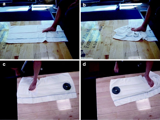

Fig. 18.2
Towel curls for toe (a) flexion and (b) extension; (c, d) Towel “swishes” for ankle inversion/eversion
Formalized physical therapy visits are initiated generally from 2 to 12 weeks post-surgery with an evaluation by a physical therapist.4–12 In the United States, patients are typically seen 2–3 times/week. A detailed assessment is performed by the physical therapist including history of the injury and surgical procedure, which regions should be protected and not be mobilized (such as with arthrodesis procedures), location of hardware or implants, and generalized situation of the patient. Note should be taken as to timing of protection with boots and braces. An appreciation for the stage of the healing must be understood before the therapist determines the effective treatment plan. Different procedures will require strategically timed protection/pain control, while others will call for early motion (Table 18.1).
Table 18.1
Assessment by physical therapist
1. History of injury, date, and type of surgery |
2. Length of protection with boot, i.e., when can patient get out of the boot “OOB” |
3. Length of protection due to healing such as tendon, fusion site, or bone graft |
4. Location of hardware and implants |
5. Restriction of motion to be maintained such as with arthrodesis procedures |
6. Patients home, work, and activities including sports |
7. Degree of pain, swelling, warmth, color of surgical site, presence of external sutures. |
8. Disability (needs assistive aids), aggravating and easing factors |
9. Patient’s current amount of activity, icing, and exercising |
10. Patient’s goals/sports/occupation |
11. Current functional status: ROM, strength, gait, balance, and proprioception |
12. Surgeon’s expected return to activity “RTA” for procedure |
13. Consider objective foot scoring device (i.e., American Orthopedic Foot and Ankle Society, Foot and Ankle Assessment Module, Maryland Foot Score) |
Modalities used postoperatively (such as ultrasound, phono- and iontophoresis, and interferential current) have not been critically studied, and therefore not often used post-surgically in our clinics. Though use of electrical stimulation and ultrasound is common and may be helpful, (for swelling and adhesions, respectively) firmly established protocols have not been performed. Postoperative edema reduction can be enhanced by ice compression devices such as the GameReady™ and Aircast Cryo Cuff™ in a controlled setting (Fig. 18.3).2 Taping techniques such as Kinesio™ tape for edema control and ankle protection can be helpful (Fig. 18.4). Lymph massage has been proven as helpful and is utilized as needed. This is particularly helpful in patients having multiple surgeries, incisions, and venous stasis.10
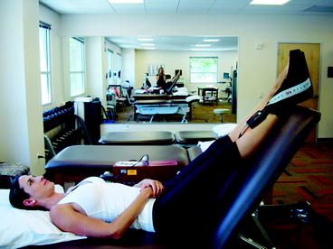
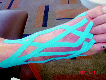

Fig. 18.3
Game Ready™ ice device and elevation

Fig. 18.4
Kinesio™ tape for ankle swelling
Ergonomic equipment such as stationary bikes and elliptical trainers is utilized at appropriate times. Strengthening programs have primarily been studied post-ankle sprain and for nonsurgical treatment of Achilles tendonopathy often, with good success. The eccentric programs for Achilles tendonopathy have not been studied post-surgery. We therefore initiate rehabilitation with concentric strengthening programs post-surgery.7,8,13,14 Eccentric exercises can be utilized in the later phases of rehabilitation after gait is normalized.15–18 We also recommend hip stabilization exercises as recent studies point to hip weakness as a factor in ankle injuries.18–20
A recent device we have found helpful for lower extremity strengthening and rehabilitation is the “Alter-G™ Treadmill” (Alter-G, Milpitas, CA USA) (Fig. 18.5). This specialized treadmill allows adjustment and reduction of bodyweight such that if a patient is unable to perform lower extremity tasks such as heel raises or walking, the machine can be calibrated to allow them to do so. In our clinic, we typically reduce the body weight 30–50% and initiate single-legged concentric heel raises and restore normal gait (Table 18.2).8
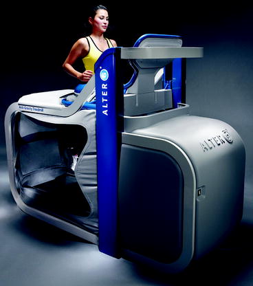

Fig. 18.5
Runner on Alter-G™ treadmill
Table 18.2
Concentric heel raise progression (on successive days if pain-free):
3 sets of 10 repetitions single-legged, pain-free |
4 sets of 10 repetitions single-legged, pain-free |
5 sets of 10 repetitions single-legged, pain-free |
3 sets of 15 repetitions single-legged, pain-free |
4 sets of 15 repetitions single-legged, pain-free |
5 sets of 15 repetitions single-legged, pain-free |
3 sets of 20 repetitions single-legged, pain-free |
4 sets of 20 repetitions single-legged, pain-free |
5 sets of 20 repetitions single-legged, pain-free |
3 sets of 25 repetitions single-legged, pain-free |
4 sets of 25 repetitions single-legged, pain-free |
5 sets of 25 repetitions single-legged, pain-free |
18.1 Rehabilitation Programs Used for Ankle Procedures
This program is followed after ankle procedures, i.e., Achilles, posterior tibial tendon reconstruction procedures, peroneal tendon repair, ankle stabilization or fracture repair, and osteochondral defect repair (with bone graft) or chondral lesion (micro-fracture).
Active ROM is allowed at 3 weeks (generally after cast removal) working on plantarflexion and inversion/eversion with a towel (Fig. 18.6). A below-knee cast boot that is maintained for immobilization is removed for exercise. Patients may be non-weight-bearing from 2 to 6 weeks depending on the procedure. Formal physical therapy is initiated between 8 and 10 weeks (“slower healing” procedures start later), though cross-training on a stationary bike is allowed with the boot/cast (with the heel on the pedal) as soon as 1 week post-surgery.6,7 Swimming is allowed (without flip-turns) between 4 and 6 weeks post-surgery. Physical therapy includes progressive strengthening with visits on a bi-weekly basis with a physical therapist. It should be noted that stretching, thought to be appropriate post-injury, may have a limited role post-surgery. It must be done under proper supervision.14,21
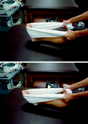

Fig. 18.6
(a, b) Plantarflexion exercises with towel
18.1.1 Phase 1
18.1.1.1 Weeks 8–10 Post-surgery
Initial evaluation;
Protected mobilization, i.e., dorsiflexion to tolerance with Achilles procedures, no eversion beyond neutral with posterior tibial/medial stabilization, no inversion beyond neutral with peroneal/lateral stabilization procedures. Also, note, no mobilization of fused joints; (Figs. 18.7 and 18.8).
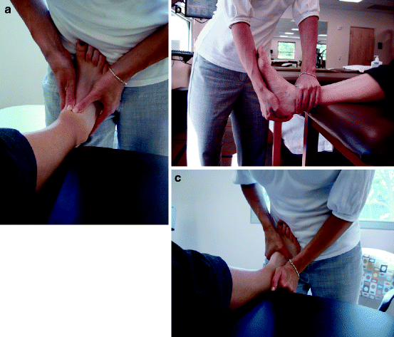
Fig. 18.7
(a–c) Distraction with anterior and posterior “glides” (mobilization) of the subtalar joint
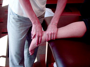
Fig. 18.8
Mobilization (PA glide) of the subtalar joint
Non-weight-bearing ankle strengthening with surgical tubing (inversion, eversion, dorsiflexion and plantarflexion) (Fig. 18.9).

Fig. 18.9
(a) Ankle/STJ inversion (right foot) and (b) Ankle/STJ eversion (right foot) with elastic tubing
Cross-friction massage to incision (once fully healed) and posterior ankle (Fig. 18.10).
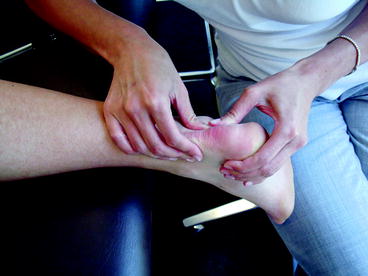
Fig. 18.10
Cross-friction massage of ankle tendons
Seated and standing calf stretch (Fig. 18.11).
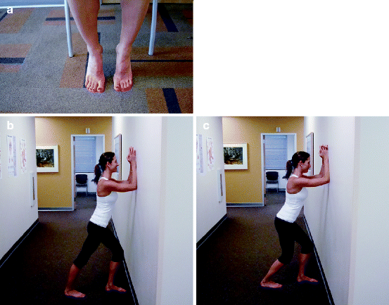
Fig. 18.11
Seated (a) and standing with knee straight (b) and knee bent (c) calf stretch
Introduce single-limb proprioception (Fig. 18.12).
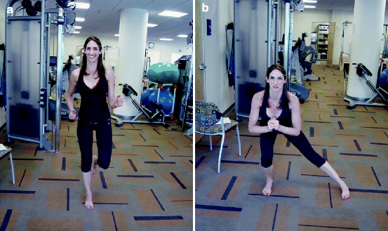
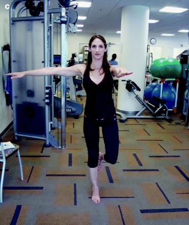
Fig. 18.12
(a–c) Single leg, side squat/lateral lunge, back to single leg ankle stability exercise on flat surface, for gluteal strengthening
Bilateral concentric heel raises (can start in a pool, Alter-G™ or on leg press machine if patient is unable to do it on flat ground) (Fig. 18.13).
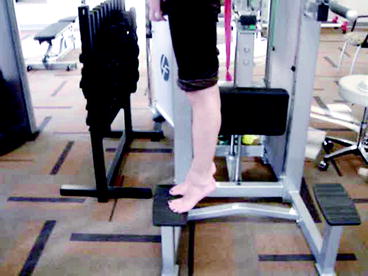
Fig. 18.13
Bilateral concentric heel raise
Home instruction on strengthening with a towel.
Cryotherapy for 15 min at session end.
18.1.1.2 Weeks 10–12+ Post-surgery
Same as above, plus:
Get Clinical Tree app for offline access

Soft-tissue massage to calf muscle and posterior ankle tendons (Figs. 18.14 and 18.15)
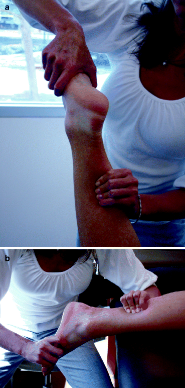
Fig. 18.14
(a, b) Mobilization of ankle
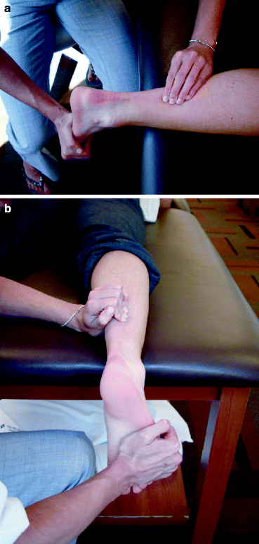
Fig. 18.15
(a, b) Mobilization of calf
Stay updated, free articles. Join our Telegram channel

Full access? Get Clinical Tree


