Stage
Definition
1
The elbow subluxates in a posterolateral rotatory direction and will have a positive lateral pivot shit test due to lateral ulnar collateral ligament disruption
2
The elbow incompletely dislocates and the coronoid becomes perched under the trochlea due to all lateral based structures being disrupted including some anterior and posterior capsule involvement
3
Elbow completely dislocates so that coronoid is behind humerus and is due to lateral and medial sided disruption
3a
Anterior band of the medial collateral ligament (MCL) remains intact after dislocation and reduction and elbow is stable to valgus stress
3b
Anterior band of the MCL is disrupted and after reduction the elbow is unstable to valgus stress
3c
Completely stripped ligaments and soft tissue of the elbow remain unstable after reduction and splinting
21.4 Patient History and Symptoms
Most commonly, the patient will present with nonspecific pain and clicking about the elbow. While the traumatic dislocation may produce obvious instability, it is much more common for the athlete to present with a more subtle form of instability, due to multiple small injuries. The injured athlete with these more subtle patterns will not give a clear history of a precipitating event, but rather will, more commonly, describe a gradual onset with slow worsening of lateral elbow pain. When O’Driscoll first described this condition, his patients presented with symptoms consistent with recurrent dislocation of the elbow or of the proximal radioulnar joint [1]. Many patients report a history of an elbow dislocation. Almost half of elbow dislocations occurred in sport, including basketball, football, and wrestling in males and gymnastics and skating in females [19].
Patients who partake in racquet sports such as tennis, squash, and racquetball tend to present with signs and symptoms of lateral epicondylitis (tennis elbow); however, a small cohort of patients will injure their LCL complex displaying symptoms of PLRI [19]. Patients with a history of traumatic elbow injury treated with radial head excision will also display symptoms of PLRI [21].
Patients tend to report instability symptoms when the elbow is extended and the forearm is supinated [1]. Other common complaints include pain, giving way, locking, clicking, or snapping of the elbow [1, 2, 4]. These symptoms usually present with the elbow in terminal extension and with the forearm in supination [21]. A common activity patients will describe that exacerbates their symptoms will be carrying a grocery bag and that the elbow feels unstable [5]. Patients will seldom present with recurrent or complete elbow dislocations [22].
21.5 Physical Exam
Inspection of the injured elbow will often not show any abnormalities, but close visualization may reveal slight swelling along the posterolateral gutter and plica. Palpation is critical in this evaluation to discern the difference between lateral epicondylitis, radial tunnel syndrome, instability, and radiocapitellar arthritis. Sportsmen with instability will not be tender over the radial tunnel and lateral epicondyle but more posteriorly along the back of the radiocapitellar joint. Limited pronation and supination with lateral compression will not be painful, but when valgus force and increased supination in about 100° of flexion is added, the patient will often be tender along the proximal end of the radial ulnohumeral ligament (RUHL) complex and also have increased feelings of instability (Fig. 21.1).
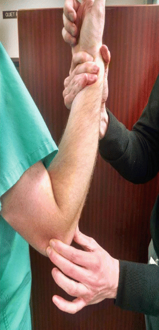
Fig. 21.1
Diagnostic maneuver for PLRI, demonstrating a valgus force applied to a fully supinated forearm at 100° of flexion
O’Driscoll [1] first described the lateral pivot shift test for PLRI. He describes testing with the extremity above the head of the patient, with the shoulder externally rotated. The test begins with the patient’s forearm fully supinated; the physician then grasps the wrist and flexes the elbow from extension while applying valgus, supinating, and axial forces to the extremity. If PLRI exists, at approximately 40° of flexion, the dislocated radiohumeral joint will be most visible as a posterior prominence and a skin dimple proximal to the radial head. Continued flexion will result in reduction of the joint, and the dimple should disappear. This test is most accurate under anesthesia (Figs. 21.2 and 21.3); however, patients will present with apprehension when awake from the procedure [1].
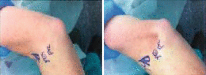
Figs. 21.2 and 21.3
Before and after pictures of the elbow prior to the lateral pivot shift test, demonstrating the dimpling of the skin at the radiohumeral joint
In most patients, the O’Driscoll test cannot be performed while awake, so our institution uses a modified test where we palpate the posterior radiocapitellar joint while more gently supinating the forearm and providing a slight valgus stress. In extension, there is no shifting or pain, but as the elbow is flexed to 90–110°, the radial head can be felt to shift posteriorly and away from the humerus.
Other tests have been determined from further research into this topic that provides physicians a wide array of exam maneuvers to help provide diagnosis for their patients [4, 15] (Table 21.2).
Table 21.2
Diagnostic physical exam maneuvers for PLRI
Test | Physical exam procedure |
|---|---|
Posterolateral rotatory drawer | Pull posteriorly on the lateral side of the proximal forearm. Positive result signified by presence of dimple or apprehension |
Table top relocation rest | Patient begins with one arm, forearm in supination and presses up on table. Test positive if apprehension at 40° of flexion, and patient relieved if physician presses on radial head |
Prone push-up | Patient begins with elbows flexed at 90°, arms abducted, and forearms supinated lying prone on floor. Positive result with dimple or apprehension upon push-up |
Chair push-up | Patient begins with elbows flexed at 90°, arms abducted, and forearms supinated seated in chair. While patient pushes down on chair, test is positive if dimple or apprehension |
21.6 Imaging
The initial work-up for LCL injury imaging should begin with anteroposterior and lateral radiographs of the elbow. Radiographs of patients’ elbows consistent with LCL disruption may show some posterior displacement of the radial head relative to the capitellum or a drop sign (slight widening of the ulnohumeral joint, >3 mm) [4, 19]. Other times a small avulsion fragment off of the lateral epicondyle may be present; however, most patients with PLRI present with negative plain radiographs [23] (Fig. 21.4). Stress films may also be obtained, including anteroposterior films with varus loading. A positive finding would show gapping at the lateral joint of the elbow [23]. A stress lateral can be performed with the forearm maximally supinated and a lateral pivot shift being performed. A positive finding would show a widened ulnohumeral joint and an inferiorly subluxated radial head [23].
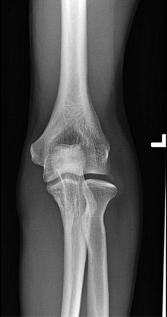
Fig. 21.4
Anterior posterior (AP) radiograph of the elbow, showing an avulsion fracture of the lateral epicondyle consistent with an LCL complex injury
Magnetic resonance imaging (MRI) may be used to further evaluate the joint in question. Coronal imaging, slices that are <2 mm slices, provides the best visualization of the collateral ligament complexes [11] (Fig. 21.5). In younger patient populations, the LUCL will appear striated on imaging [24]. MRI tends to show LCL tears at the proximal origin of the humeral condyle. If the patient has PLRI, there can be posterior displacement of the ulna and radial head in regard to the humerus, and this has been coined as “perched elbow” [19]. It is recommended to detect ulnar lateral collateral ligament abnormalities that MRI is obtained with three-dimensional gradient-echo and fast-spin-echo sequences [11]. The addition of contrast can aid in the diagnosis [25, 26]. LCL injuries will present as ligament redundancy, attenuation, or discontinuity on imaging [11]. An avulsion fracture may also be seen at the proximal ligamentous attachment or underlying marrow edema in the humerus [27].
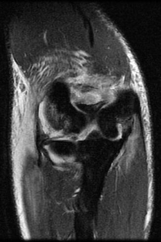
Fig. 21.5
Coronal MRI showing disruption of the LCL complex from the lateral epicondyle to the radius
In most cases, the magnetic resonance arthrogram represents the most definitive test.
21.7 Treatment
21.7.1 Nonoperative Management
In more limited instability cases, a compressive sleeve and strengthening of the lateral musculature may be effective. This treatment may be supplemented by topical nonsteroidal anti-inflammatory drugs (NSAIDs) and physiotherapy treatments that allow the athlete to return to sport. Although an attractive option, we were unable to uncover reports of ligament regeneration utilizing biologic supplementation.
21.7.2 Operative Management
The most important is identifying the patients who would benefit from surgery. These include those that are symptomatic with pain, signs of instability, or restrictions in their daily lifestyle [28]. Described are several techniques that have reported success in treating patients.
21.7.2.1 Arthroscopic Repair
Arthroscopic repair is performed in the prone position with the arm stabilized on a bolster. The portals used for accessing the joint include the proximal anteromedial and proximal anterolateral portals for the anterior compartment and the posterior central and posterolateral portals for the posterior compartment.
For an acute ligament rupture, the anterior compartment is accessed first, and a diagnostic arthroscopy is performed. Any hematoma can be evacuated at this time with an arthroscopic shaver. Upon complete anterior evaluation, the arthroscope is placed into the posterior compartment for further diagnostic purposes. At this point in the procedure, it is noted that in an elbow with LCL instability, the arthroscope is able to be placed down the posterolateral gutter across the ulnohumeral articulation into the medial gutter. This is called the “drive-through sign of the elbow.” This is only possible in an unstable elbow, and once the complex is repaired, this should no longer be possible.
Related posts:


Stay updated, free articles. Join our Telegram channel

Full access? Get Clinical Tree


