, Paul D. Siney1 and Patricia A. Fleming1
(1)
The John Charnley Research Institute Wrightington Hospital, Wigan, Lancashire, UK
Position of the stem within the medullary canal in cemented total hip arthroplasties (THA) has been under scrutiny for some time. Initially this was in the context of stem fracture [1] then in relation to the predictability of the long-term outcome of stem fixation [2]. In both these publications only the antero-posterior (AP) radiographs were examined. More recently the attention has been focused on the high incidence of early failures [3] and suggestions made that endosteal cavitation and component loosening may be due to the direct effect of the ultra high molecular weight polyethylene (UHMWPE) wear particles gaining access to the bone cement interface through cement defects. As a result the trend in the design, instrumentation and the surgical technique has been to facilitate central placement of the stem within the medullary canal avoiding stem tip-cortex contact and aiming at a cement mantle of a predetermined thickness. To what extent does the position of the stem within the medullary canal in cemented THA play a part in the long-term outcome has not been established with certainty. Since a prospective study would not be possible, let alone acceptable, and a long-term follow-up would be necessary, [4] we have attempted to answer the question by examining both failed and successful cases retrospectively.
The review being retrospective it was not possible to comment on the cementing technique or the relevance of the cement mantle thickness as it was often disrupted by the process of loosening in the failed cases.
Successful result was defined as a patient who has had a cemented THA with a minimum follow-up of 8 years and considered the result to be clinically successful: the patient being free from the pre-operative hip pain, the hip having full or nearly full range of movement and the patient’s activity level being normal or near normal for age, gender and the underlying hip pathology.
Failed result was defined as a patient who has had a revision for aseptic loosening of a cemented stem.
All the relevant information was extracted from the records and antero-posterior and lateral radiographs of the hip were examined. In the failed cases referred from other units, only the pre-revision AP and lateral radiographs were available.
Definition of Stem Position
Antero-posterior Radiographs
Neutral position was defined when a line drawn through the centre of the shaft of the stem was in line with the centre of the medullary canal and an imaginary extension of the line drawn through the centre of the shaft of the stem would not encroach onto the femoral cortex as seen on the AP radiograph of the pelvis centred over the symphysis pubis (Fig. 25.1).
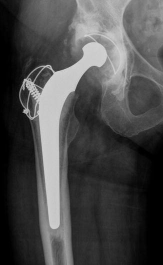
Fig. 25.1
Neutral stem position on an AP radiograph
Valgus position was defined as the stem positioned laterally, within the medullary canal, in its proximal part with its distal end towards the medial femoral cortex, irrespective of whether or not the tip was touching or even encroaching into the medial femoral cortex (Fig. 25.2).
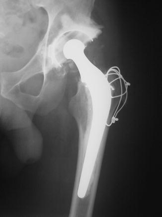
Fig. 25.2
Valgus stem position on an AP radiograph
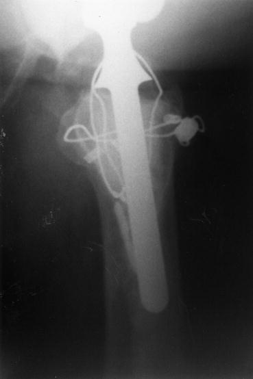
Varus position was when the stem was positioned towards the medial femoral neck proximally while its tip was directed towards the lateral femoral cortex, irrespective of whether or not it was touching or encroaching into the lateral femoral cortex (Fig. 25.3).
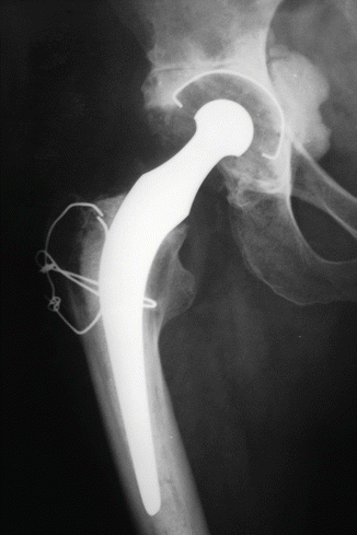
Fig. 25.3
Varus stem position on an AP radiograph
Lateral Radiographs
Neutral position as defined on the A.P. radiograph, (Fig. 25.4) but the radiograph was not centred over the symphysis pubis.
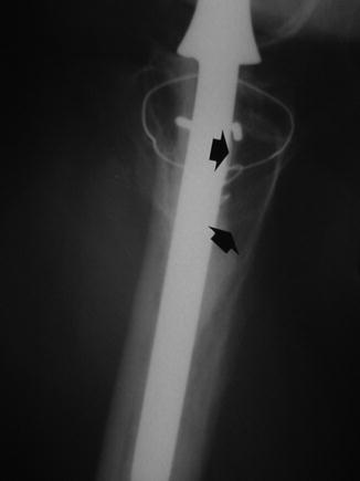
Fig. 25.4
View of lateral radiograph with stem in neutral position. Calcar femorale not cleared
Antero–posterior position was defined when the stem entered the medullary canal through or near the area of sectioned femoral neck and its tip was directed towards the posterior femoral cortex irrespective of whether it was touching or encroaching into the cortex (Fig. 25.5).
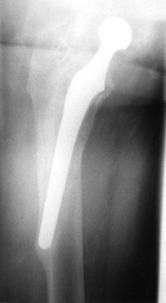
Fig. 25.5
View of lateral radiograph with stem positioned front to back
Postero–anterior position was defined when the tip of the stem was directed towards the anterior cortex. irrespective of whether it was touching or encroaching into it (Fig. 25.6).