1.
History
1. Co-morbidity
2. Etiology
Acute
Progressive
3. Symptoms
Pain
Stiffness
Locking
Paresthesia
4. Location
2.
Inspection
1. Resting position
2. Localized swelling
3. Carrying angle
4. Anatomical areas
5. General inspection
3.
Palpation
1. Lateral
2. Medial
3. Posterior
4. Anterior
4.
Passive motion
1. Flexion – extension
2. Pronation – supination
3. Stability
Valgus
Valgus stress test
Moving valgus test
Milking maneuver
Varus
Varus stress test
Posterolateral
Lateral pivot shift
Posteromedial
Medial pivot shift
5.
Active motion
1. Flexion – extension
2. Pronation – supination
6.
Active motion against resistance
1. Brachialis
2. Biceps
Hooktest
3. Triceps
4. ECRB (tenniselbow)
5. FCR + pronator teres (golfer’s elbow)
6. Stability
Posterolateral
Push-up test
Chair test
7.
Neurologic examination
1. Ulnar nerve
2. Radial nerve
3. Median nerve
8.
Lidocaine test
Table 3.2
Anatomical area and differential diagnosis
Lateral | Medial | Anterior | Posterior |
|---|---|---|---|
Radiocapitellaire artrose Osteochondrale loose body Radial head fracture Osteochondritis dissecans Tenniselbow | UCL lesion Ulnar neuritis Ulnar subluxation Golfer’s elbow | Distal bicepstendon rupture Anterior capsule strain Coronoid osteophyte formation | Valgus extension overload Posterior osteophyte with impingement Tricepstendinitis Olecranon bursitis Olecranon stress fracture |
Next, the evaluator should inquire about mechanical symptoms, such as clicking with motion, locking in extension, and catching, which can be caused by intra-articular pathology. Also loss of extension and/or flexion needs to be identified and can occur progressively. Loss of range motion can be one of the first complaints in overhead athlete.
Patient reports of numbness and tingling distal to the elbow with specific attention to the ring finger and little finger need to be evaluated. Often, these symptoms come and go.
3.2 Inspection
Inspection and observation of the elbow begins as the patient walks into the examination room or as he or she comes off the field.
Inspection of the elbow should be carried out in a systematic fashion.
When starting the inspection of the elbow, it is important to visualize both arms for comparison. The examiner should note the resting position of the painful elbow. A patient with significant joint effusion will hold the elbow at 70–80° of flexion, as this corresponds to the position of maximum volume of the elbow joint [5].
Also localized swelling should be examined. For example, swelling over the olecranon can indicate olecranon bursitis from trauma or underlying inflammation.
Next, the carrying angle is evaluated (Fig. 3.1). The normal carrying angle in full extension is approximately 11–14° of valgus in adult men and 13–16 in adult women [11]. Variations in carrying angle may be due to previous trauma, developmental abnormality, injury, or adaptive changes. This angle is greatest in valgus at full extension, diminishing during flexion, and became varus at full flexion [8].
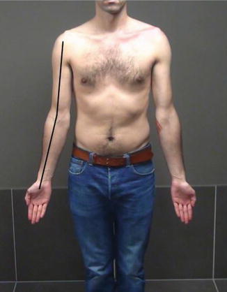
Fig. 3.1
Carrying angle
Regardless of the order of inspection, the clinician should make note of several important anatomic areas including the lateral recess, olecranon, medial epicondylar region, and antecubital fossa [5]. Also, differences seen in muscle mass may be due to injury or to hypertrophy in the dominant arm.
The clinician should complete the inspection by looking at the topographical landmarks of the entire upper extremity and trunk. Scapular winging or significant atrophy of the deltoid or rotator cuff musculature may be the cause of abnormal mechanics that result in undue stresses across the elbow articulation. This determination will permit the clinician to prescribe appropriate treatment to correct the true pathology. Similarly, the distal aspect of the extremity should be inspected to assess for discoloration of the fingers and fingertips or bony deformity.
3.3 Palpation
Palpation of the elbow should be carried out in a systematic fashion and can also be subdivided into the four anatomic regions.
First, the lateral part of the elbow with the lateral epicondyle and the radial head are palpated. Pain directly at the lateral epicondyle can be due to trauma or LCL injury. Tenderness due to lateral epicondylitis is theoretically just anterior and distal to the epicondyle at the origin of the ECRB.
When palpating the radial head, there may be tenderness and associated clicking over the radial head with rotation seen in fractures, arthrosis, or a symptomatic posterolateral synovial plica. Next, palpation of the lateral recess, or soft spot, can easily identify an elbow effusion.
At the medial side, the medial epicondyle, medial collateral ligament (MCL), and the ulnar nerve can be palpated. The MCL is palpated with the elbow in 50–70° of flexion to move the overlying medial muscles anterior to the MCL.
Related posts:


Stay updated, free articles. Join our Telegram channel

Full access? Get Clinical Tree


