Fig. 15.1
(a) Pre-op X-ray showing avulsed peroneal retinaculum. (b) MRI showing torn peroneal retinaculum. (c) Diagram of the classification of peroneal retinaculum tears. PLT, peroneus longus tendon; PBT, peroneus brevis tendon; SPR, superior peroneal retinaculum
15.1.5 Surgical Techniques
Many surgical techniques limited only to case series have been described but only Level IV/Grade C evidence has been produced. No randomized studies have been conducted to determine which procedure is the most successful. (Table 15.1)38–56 Generally, five categories of surgical repair are listed: (1) Anatomic reattachment of the retinaculum; (2) Reinforcement of the superior peroneal retinaculum with local tissue transfers; (3) Rerouting the peroneal tendons behind the calcaneofibular ligament; (4) Bone block procedures; (5) Groove-deepening procedures.
Table 15.1
Studies and surgical techniques for the management of peroneal tendons subluxation
Authors | Year | Level of evidence | Number of cases | Procedures |
|---|---|---|---|---|
Eckert and Davis25 | 1976 | IV | 73 | Anatomical reattachment of SPR |
Marti38 | 1977 | IV | 12 | Modified Kelly |
Zoellner and Clancy12 | 1979 | IV | 9 | Groove deepening |
Escalas et al.4 | 1980 | IV | 28 | Jones procedure |
Arrowsmith et al.39 | 1983 | IV | 6 | Anatomical reattachment of SPR plus groove deepening |
Poll and Duijfjes40 | 1984 | IV | 10 | Rerouting tendon under CFL |
Martens et al.41 | 1986 | IV | 11 | Rerouting tendon under CFL |
Micheli et al.42 | 1989 | IV | 12 | Modified kelly |
Orthner et al.43 | 1989 | IV | 23 | Anatomical reattachment plus screw |
Wirth44 | 1990 | IV | 15 | Modified Viernstein and Kelly |
Thomas et al.45 | 1992 | IV | 31 | Modified Ellis-Jones |
Steinböck et al.46 | 1994 | IV | 13 | Rerouting tendon under CFL |
Mason and Henderson47 | 1996 | IV | 11 | Anatomical reattachment of SPR |
Karlsson et al.48 | 1996 | IV | 15 | Soft tissue reconstruction of superior retinaculum |
Kollias and Ferkel49 | 1997 | IV | 12 | Groove deepening |
Hui et al.50 | 1998 | IV | 21 | Anatomical reattachment of SPR |
Mendicino et al.51 | 2001 | IV | ? | Groove deepening |
Shawen et al.52 | 2004 | IV | 20 | Groove deepening |
Porter D, McCarrol J et al.53 | 2005 | IV | 13 | Groove deepening |
Adachi et al.54 | 2006 | IV | 20 | Anatomical reattachment of SPR |
Maffulli et al.55 | 2006 | IV | 14 | Anatomical reattachment of SPR |
Ogawa et al.56 | 2007 | IV | 15 | Groove deepening |
15.1.5.1 Anatomic Reattachment of SPR
The aim of anatomic reattachment of the SPR is the restoration of the primary restraint of the peroneal tendons. Reattachment with sutures brought through drill holes in the distal fibula has been described by several authors.8,25,38–40,47,48 Alternatively, Beck6 brought the retinaculum through a slip produced in the distal fibula and fixed this with a screw, reporting on nine patients without complication. Eighteen of 21 patients treated with the “Singapore operation” at 9 years had excellent results. Three patients experienced postoperative pain and neuromas, but no recurrence was noted.50 Karlsson and colleagues reported 13 patients with good to excellent results associating a groove deepening in conjunction with reattachment of the SPR if the posterior surface of the fibula was flat or convex.48 Orthner et al. obtained excellent results for peroneal tendons subluxation suturing side by side the SPR in acute lesions.43 Adachi et al. proposed retinaculoplasty, opening the false pouch through one incision, and suturing the SPR to the fibula while tensioning it. In this study, the authors reported that 15 of the 18 patients involved in sports activities returned to their previous activities without reducing their activity levels.54 Recently, an endoscopic technique for the anatomical repair of the SPR has been described.57 Anatomic reattachment of superior retinaculum seems to be the preferred technique in patients with acute subluxation of peroneal tendons.
15.1.5.2 Reinforcement of SPR with Soft Tissue
Several authors have described procedures to augment or reinforce an attenuated SPR with soft tissue transfer. Ellis-Jones58 first described restraining the peroneal tendons with a strip of Achilles tendon anchored through a drill hole in the fibula. No recurrences were noted in a long-term follow-up of 15 patients who underwent the Ellis-Jones repair.4 Thomas et al.45 described a modification to this procedure that allowed the use of a smaller strip of Achilles tendon, reducing the risk of weakening the Achilles tendon. Use of the tendon of peroneus brevis,39,59,60 plantaris61,62 and peroneus quartus63 have been described for the same purpose. Zoellner and Clancy12 and Gould9 used periosteal flaps to restrain the peroneal tendons in a deepened peroneal groove with satisfactory results. In patients treated with a periosteal flap from the retrofibular groove on its own or together with groove deepening, no postoperative complications were noted.64
15.1.5.3 Rerouting the Peroneal Tendons Behind the Calcaneofibular Ligament
This surgical technique does not address the issue of restoration of the anatomy of the SPR, but uses the calcaneofibular ligament as the natural alternative restraint. Platzgummer65 and later Steinbock et al.46 divided the calcaneofibular ligament, transposed the tendons behind it, and sutured the calcaneofibular ligament back together. The 13 patients operated with this technique showed good or excellent results, with no evidence of recurrence or instability. Sarmiento and Wolf divided the peroneal tendons and re-sutured them after rerouting them behind the calcaneofibular ligament66; 11 patients showed no evidence of recurrence or instability at follow-up, although two patients suffered a sural nerve injury. Martens et al. used the same technique of Sarmiento with excellent results at 30-month follow-up in 11 patients.41 Both methods may potentially weaken the relevant structures. To preserve the integrity of the calcaneofibular ligament, a bone block of the ligamentous insertion on the fibula67 or the calcaneus40 can be mobilized, the tendons are transposed, and the bone block is reattached with a screw. Pozzo and Jackson11 reported no complication and return to full level of activity in a case report. Poll and Duijfjes40 reported ten patients with no recurrence or instability. Ferroudji et al. reported their experience with 19 patients, with excellent results in 17 patients. Sports activities were resumed after an average of 3.3 months.68 This surgical technique should be preferred in patients with chronic luxation of the peroneal tendons in whom the SPR is absent.
15.1.5.4 Bone Block Procedure
These surgical procedures were developed to deepen the retrofibular groove using a bone graft as a physical restraint to the peroneal tendons. In 1920, Kelly67 described a bone block procedure using screw fixation and later designed a wedge-shaped graft that avoided the use of screws near the ankle joint. Watson-Jones and DuVries69,70 modified Kelly’s technique. Watson-Jones69 used an osteoperiosteal flap anchored by a soft tissue pedicle, and secured it posteriorly with sutures. DuVries70 anchored a posteriorly displaced wedge with a screw. Other authors reported on patients with chronic subluxation operated with a modified Kelly technique with no recurrence.38,44 In 1989, Micheli and colleagues42 treated 12 patients with an inferiorly displaced fibula bone graft fixed with screws; one patient suffered a traumatic fracture of the graft, and two required exploration for pain; there were no recurrences of the subluxation. Adhesion of the peroneal tendons to the fresh bone wound, fractures of bone grafts, and the need for metalwork are major disadvantages of bone block procedures.6 This surgical technique seems to be the more exposed to intraoperative and postoperative complications, and should be reserved for selected cases.
15.1.5.5 Groove Deepening
Patients presenting with a flat or convex retrofibular sulcus could be managed with this surgical technique. Zoellner and Clancy12 elevated an osteoperiosteal flap on the posterior aspect of the distal fibula, and removed cancellous bone with a gouge. The flap was then reduced into the deepened sulcus, and the tendons replaced into this. Their nine patients had excellent results with no recurrence or instability. Hutchinson and Gustafson described a similar method in combination with SPR reattachment. Of 20 patients, three had poor results with recurrence of the subluxation, and one of these developed reflex sympathetic dystrophy.71 Gould9 reported a single patient in whom groove deepening was incorporated with restraint of the peroneal tendons by reflection of elevated osteoperiosteal flap. Recently, Ogawa et al. used an indirect fibular groove–deepening technique.56 Mendicino and colleagues51 employed intramedullary drilling and cortical impaction to achieve groove deepening. Porter et al. proposed groove deepening associated with an accelerated rehabilitation program. Eight of 13 patients returned to pre-injury sports participation.53 The depth of the retrofibular sulcus was previously thought to play an important role in restraining the peroneal tendons.49,52 Recently, however, the need for groove deepening has been questioned. Anatomic studies demonstrate the incidence of a flat or convex sulcus as high as 18%,72 28%,58 and 30%.73 The low incidence of peroneal tendon subluxation would suggest that the morphology of the groove is not a predisposing factor to subluxation.72 Histologic studies demonstrating that the peroneal groove is defined by the fibrocartilagenous periosteal cushion and not by the bony sulcus add weight to this argument.
15.1.6 Preferred Surgical Technique
Under general or spinal anesthetic, the patient is placed supine on the operating table with a sandbag under the buttock of the operative side to internally rotate the affected leg. A tourniquet is applied to the thigh, the leg exsanguinated, and the cuff inflated to 250 mmHg. A 5-cm longitudinal incision is made along the course of the peroneal tendons. The incision starts posterior to the tip of the lateral malleolus and progressed proximally, staying well anterior to the sural nerve. The incision is deepened to the peroneal tendon sheath, which is incised longitudinally 3 mm posterior to the posterior border of the fibula. Normally, the SPR itself is thin and deficient, and it was detached from its posterior attachment on the calcaneus in our case series.55 The peroneal tendons are identified by blunt dissection and protected. Attrition lesions and longitudinal tears of peroneal tendons when found are treated with a very gentle débridement or suturing with absorbable sutures.74–76 After that, we expose the lateral aspect of the lateral malleolus, and the “pouch” formed between the bony surface of the lateral malleolus and the superior peroneal retinaculum, where the tendons subluxate, becomes visible. The SPR does not heal back to its normal attachment on the posterolateral aspect of the fibula but in an elongated fashion more anteriorly on the lateral aspect of the fibula, creating a pouch on the lateral fibula into which the tendons can subluxate. The bony surface of the lateral malleolus is roughened with a periosteal elevator to produce a bleeding surface, and three or four anchors (Mitek GII, Ethicon Ltd, Edinburgh, Scotland) with 2/0 absorbable sutures (Vicryl, polyglactin 910 braided absorbable suture, Ethicon) are inserted along the posterior border of the lower fibula (Fig. 15.2). After manual testing that the anchors cannot be dislodged, the SPR is reconstructed in a “vest over pants” fashion,74 making sure that the pouch between the bony surface of the lateral malleolus and the SPR is totally obliterated. The ankle is kept in eversion and slight dorsiflexion so that the peroneal tendons were in the “worst possible position.” The strength of the repair is tested moving the ankle through the whole range of motion. The wound is closed in layers with 2/0 Vicryl for the subcutaneous fat, un-dyed 3/0 Vicryl for subcuticular, and Steri-Strips for the skin (3M, Loughborough, United Kingdom). Dressing swabs, dressing, and crepe bandage are applied. A below-the-knee walking cast is applied with the ankle in neutral and slight eversion. Weight bearing is allowed from the day after the operation, and the cast is removed 4 weeks after the procedure, when rehabilitation is started. Gradual return to activities and to sport is allowed during the course of 3–4 months from the procedure.55,76
Fig. 15.2
Anatomical reattachment of the superior retinaculum with anchors. (a) Diagram and (b) X-ray of anchor placement. (c) Intraoperative view of suture placement for retinaculum repair
15.1.7 Postoperative Care
Patients are discharged the day after surgery, after having been taught to use crutches by an orthopedic physiotherapist. No thrombo-prophylaxis is used. Patients are allowed to bear weight on the operated leg as tolerated, but are told to keep the leg elevated as much as possible for the first 2 postoperative weeks. Patients are seen on an outpatient basis at the second postoperative week, and the cast is removed 4 weeks from the operation. Patients then mobilize the ankle with physiotherapy guidance. They are allowed to partially weight-bear, and commenced gradual stretching and strengthening exercises over 8–10 weeks after surgery. Cycling and swimming are started 2 weeks after removal of the cast. Patients are allowed to return to their sport on the fifth postoperative month.55,75,76
15.2 Surgical Techniques for Peroneal Tendon Tears
15.2.1 Introduction
Peroneal tendon tears were thought to be uncommon and a relatively new entity when reported by Evans in 1966. Peroneal tendon tears were seldom described until the early 1990s, when numerous case reports and moderately sized case series were published.23,77–89 Peroneal tendon tears are thought to occur from both acute trauma and chronic instability, with and without subluxation. Peroneal retinaculum insufficiency is described in the preceding section. Chronic retinaculum insufficiency has been associated with peroneal tendon tears.80–82,90,91 As with peroneal retinaculum pathology, tendon tears can often be difficult to recognize immediately. Diagnostic tests and clinical exam are paramount. Sobel and Mizel describe the proximal injuries as “Zone I” and the distal injuries associated with the Os Peroneum as “Zone II.” Zone I injuries generally involve tears and dislocations of the tendon of peroneus brevis, while Zone II injuries involve Peroneus Longus pathology often with an Os Peroneum. They coined this condition POPS, Painful Os Peroneum Syndrome.81 Surgical treatment for both regions is often effective; the current treatment recommendations are based on Level IV & V Evidence.80
15.2.2 Anatomy
Much of the pertinent anatomy has been described in the previous section. In addition, the incidence of the Os Peroneum and its association with lateral foot pain should be considered.79,81 The Os Peroneum is present in 4–26% of individuals, depending on race and ethnicity.79,92–94 A low-lying muscle from Peroneus Brevis or an anomalous Peroneus Quartus has also been associated with peroneal tendon tears (Fig. 15.3).16,95,96 Peroneal tubercle hypertrophy has also been noted with peroneal tendon tears.78–81,89
Fig. 15.3
(a) MRI showing accessory peroneal (quartus) tendon and peroneal brevis tear. (b) Intra-op view showing accessory peroneal (quartus) tendon and peroneal brevis tear
15.2.3 Clinical Findings
Patients with peroneal tendon tears complain of lateral ankle and foot pain. Acute onset of pain can occur with inversion injuries. Patients may state they felt a “pop” and have ankle weakness. Ankle instability was noted as early as 1979 with peroneal tears.97 Clinically, patients may have noticeable pain with active resistance to the Peronei, and with single-legged weight bearing. Proprioception is altered. Swelling is often present along the peroneal tendons. Cavo-varus foot structure may be present causing a supinated foot structure, thereby putting more strain on the Peroneals. A Coleman block test should be performed to evaluate for a rigidly plantarflexed first ray causing hindfoot varus, versus a calcaneus varus (Fig. 15.4).80 Weight-bearing plain radiographs should identify ankle varus, degenerative arthritis, accessory ossicles, and exostoses (Fig. 15.5). An Os Peroneum is often present within the Peroneus Longus tendon in either a bony or cartilaginous form. A proximally migrated Os Peroneum indicates rupture of the Peroneus Longus (Fig. 15.6).80–82,86,88,89,94 Other bony structures can be associated with peroneal tendinopathy. A hypertrophied peroneal tubercle on the lateral calcaneus can often be associated with fraying and even laceration of the adjacent tendons, which is best visualized on MRI (Fig. 15.7).80–82,86 Subluxation of the Peronei from posterior to the fibula may result in tearing of the Peroneus Brevis in particular, more commonly in older patients (Fig. 15.8). This may be due to long-standing insufficiency.80,98 Inspection of shoe gear, inserts, and orthoses should also be performed. History of inflammatory arthritis such as rheumatoid arthritis, Gout and Reiter’s syndrome, and subsequent testing when indicated should be considered.99
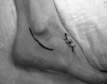
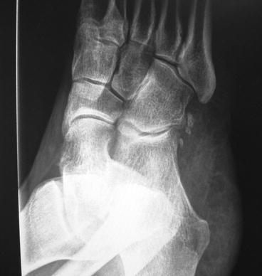
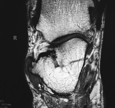
Fig. 15.4
(a) Patient with right-sided peroneus longus tear, with previous repair on left. Note plantarflexed 1st metatarsal. (b) Coleman block test showing varus rearfoot when forefoot is on the ground. (c) With forefoot suspended off the ground, the rearfoot moves to neutral. This demonstrates that the patient’s rearfoot varus deformity is due to the plantarflexed 1st metatarsal
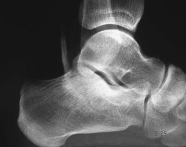
Fig. 15.5
Prominent peroneal tubercle
Fig. 15.6
Proximally migrated os peroneum indicating peroneus longus rupture
Fig. 15.7
MRI showing hypertrophic peroneal tubercle causing fraying of peroneals
Fig. 15.8
MRI showing avulsed peroneal collangenous lip (a), split peroneus brevis (b), absent peroneus brevis with empty sheath (c)
Radiographic tests for peroneal tendinopathy also include tenograms and MRI examinations. Tenograms are useful for diagnosing stenosing tenosynovitis (Fig. 15.9). MRI can also indentify this, particularly since fluid is often evident in pathological states of tendons.27,90 Ultrasound may be used as it shows fluid within the tendon sheath and has high specificity for dislocations and tears, but osteochondral and transchondral defects and accessory ossicles may not be seen with ultrasound.37,80 Fluid in the peroneal sheath may be indicative of a longitudinal tear, particularly with MRI (Fig. 15.10).80,86,88,90 MRI has a fairly high sensitivity and specificity rate, and has become the “gold standard,” though false-positives occur very commonly.80,86,90 The fact that many patients have asymptomatic tears noted on MRI exams points to the critical importance of the clinical exam.