Periprosthetic Joint Infections: Surgical Technique Two-Stage Exchange
Ajay Malviya
Fares S. Haddad
Introduction
Prosthetic joint infection (PJI) is a catastrophic complication after total hip arthroplasty (THA), with a reported incidence of less than 1% (1,2,3). The 8th Annual National Joint Registry reports that in the year 2010, in England and Wales alone, 1,055 (13% of all revisions) revision hip arthroplasties were performed for infection, with 213 of these (3% of all revisions) being single-stage procedures (4). In the United States between 1990 and 2002 there was a 50% increase in the number being undertaken (5). It has been projected that the demand for hip revision procedure would increase 174% in the next 25 years (6). The burden of prosthetic hip infections is likely to increase with the number of both primary and revision hip replacements. Significant morbidity is associated with prosthetic hip infections, including the need for further operative procedures, long-term antibiotic therapy, and prolonged hospitalization. The mortality rate from PJI is estimated to be between 1% and 2.7% (7,8,9). This is in addition to considerable financial cost to the health system. It has been estimated that in the United States the hospital costs are $96,166 per patient requiring revision arthroplasty for infection, which is 4.8 times the cost of a primary arthroplasty (10). In the United Kingdom the mean cost of revision hip replacement for deep infection has been estimated to be £21,937 per patient, which is almost twice the cost of revision for aseptic loosening (11). Similarly, reports from Australia show that the cost of managing PJI was 3.1 times more than the cost of primary joint arthroplasty (12).
When early infections occur, within 4 to 6 weeks of implantation, the implant can be left in place with a high probability of cure with early aggressive debridement whereas late infections, defined as those presenting more than 4 to 6 weeks from surgery, require revision of the prosthesis to eradicate the infection (13,14,15). In such cases, one can differentiate between one-stage and two-stage revisions. In the former a new prosthesis is implanted immediately after the removal of all foreign material in one operation. Two-stage revision involves an initial operation to remove all foreign materials either left as a Girdlestone excision arthroplasty or with the implantation of a cement spacer followed by an interim phase of 6 to 10 weeks of parenteral antibiotics, followed by reimplantation of the definitive prosthesis.
Considerable variation exists in the contemporary practices and preference of surgeons dealing with these complex cases. Treatment options for chronic hip joint infections after THA have evolved from a single-stage direct exchange to two-stage and if required multistage revision arthroplasty. The dilemma of identifying which patients are suitable for single-stage versus multistage revision remains unresolved.
While single-stage revision has had good results (16,17,18), two-stage reimplantation remains the gold standard (19) for the treatment of chronically infected THA as the successful eradication of infection is well over 90% (2,20,21,22,23). Furthermore, it allows uncemented reconstruction with the use of allografts at the second stage, which is particularly important given the frequency of femoral and acetabular defects associated with THA infections (24,25,26). The Norwegian Arthroplasty Register reveals that the survival after revision of infected primary THA with two-stage implant exchange was slightly superior to that for one-stage exchange of the whole prosthesis, despite the two-stage procedure often being used for more severe infections (27). Debridement with exchange of head and/or liner but with retention of the fixed implant (minor revision) had a 76% chance of not being re-revised within the first 2 years (27). Long-term suppressive antibiotics and salvage procedures such as Girdlestone arthroplasty, arthrodesis, and amputation have also been used in high-risk operative patients as well as those unwilling to have additional procedures (9,28,29).
Classification
Zimmerli et al. classified arthroplasty infections as follows: early (developing in the first 3 months after surgery), delayed (occurring 3 to 24 months after surgery), and late (greater than 24 months).
Toms et al. (14) have proposed a modification of the classification proposed by Fitzgerald et al. (1,30); with stage I infections presenting acutely (within the first 6 weeks), stage IIa delayed presentation with a chronic indolent infection regardless of when it presents, and stage III describing those that occur suddenly, with an acute presentation of infection secondary to hematogenous spread in an otherwise well-functioning hip replacement. Tsukayama et al. (31) has added a fourth type when a positive culture is found at the time of revision without previous evidence of infection.
These classifications roughly correlate to the observed differences in the causative pathogens; with virulent organisms such as Staphylococcus aureus characteristically presenting earlier and more indolent pathogens such as coagulase-negative Staphylococcus usually presenting later (9).
Indications for Two-Staged Revision
Two-staged revision hip replacement is considered the treatment of choice for chronically infected total hip replacements (14). Patients who have failed debridement and retention of prosthesis or those with recurrent infection after single-stage revision surgery, would be suitable candidates (2).
Contraindications for Two-Stage Revision
The main contraindication of the two-staged procedure would be patients who are deemed medically unfit to undergo the rigors of two major surgeries with the possibility of limited mobility in the interim period. The other consideration as a contraindication would be inadequate bone stock and poor soft tissue cover, which would not be able to withstand the insult of two separate surgical interventions.
Case Study
A 76-year-old gentleman (Mr. WL) had presented 6 years after an uneventful primary hip replacement with a 6-month history of gradually increasing pain and discomfort in his right hip. This had deteriorated over 3 weeks and he complained of general malaise and pyrexia.
His radiographs are shown in Figure 96.1.
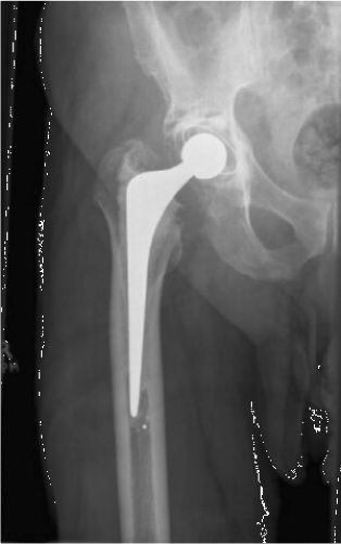 Figure 96.1. Right hip AP radiograph of Mr. WL showing circumferential radiolucent lines around both the acetabular and femoral components. |
Diagnosis
The diagnosis of infection after joint replacement relies on the surgeon’s judgment of the clinical presentation, the findings on physical examination, radiographs, and the interpretation of relevant laboratory investigations (34). In case of chronic low-grade infections the clinical diagnosis can sometimes be challenging; with many of the typical symptoms found in infection being absent. Pain is the predominant symptom of PJIs and is present in 90% to 100% of patients. The presence of fever is variable with 9% to 43% of patients in most case series having documented elevated temperatures (33,35,36,37,38). A discharging sinus is associated with chronic, indolent infection presentations (39).
Mr. WL’s blood results showed an elevated CRP of 177 mg/L, with a normal white cell count. Serologic investigations like erythrocyte sedimentation rate (ESR) and C-reactive protein (CRP) form the backbone of diagnosing infection (40,41). There are, however, limitations to their diagnostic utility. The CRP is a better indicator of infection as it is more sensitive and returns to normal within the first 3 weeks after operation, compared to the ESR, which can take up to 1 year to become normal (42). If both the ESR and CRP are elevated, the probability of infection has been noted to be 83%, and when both are negative infection may be reliably excluded (41). The search for other biochemical markers of infection has included cytokines like
interleukin-6 (IL-6) and tumor necrosis factor α (TNF-α), which are released by monocytes and macrophages in the setting of infection (40). Procalcitonin is a precursor of calcitonin and has been shown to be a specific marker of bacterial sepsis (43). High leukocyte count in the synovial fluid may assist in diagnosis of PJIs (44).
interleukin-6 (IL-6) and tumor necrosis factor α (TNF-α), which are released by monocytes and macrophages in the setting of infection (40). Procalcitonin is a precursor of calcitonin and has been shown to be a specific marker of bacterial sepsis (43). High leukocyte count in the synovial fluid may assist in diagnosis of PJIs (44).
The radiographs of WL (Fig. 96.1) showed lucency around the acetabular components with evidence of loosening. Plain radiographs lack sensitivity and specificity in diagnosing prosthetic infection with clear difficulties in distinguishing between septic and aseptic loosening. In early infection plain radiographs are frequently normal (45). Chronic infection can cause radiographic changes, including periostitis, osteopenia, endosteal reaction, and rapid progressive loosening or osteolysis (14).
Radionuclide bone scan (Technetium-Methylene Diphosphonate—99mTc) is a sensitive test for PJI, but lacks specificity, as it does not differentiate between aseptic and septic loosening (46). Indium-111–labeled white cell scans have a much higher sensitivity in infection, but is expensive and time consuming (47). Similar findings have been documented with the 18F-Fluorodeoxyglucose positron emission tomography (FDG-PET) (48,49).
Microbiologic Analysis
The identification of the causative pathogen in a prosthetic infection is critical to the institution of appropriate management, including selection of the most appropriate antibiotic to target the pathogen while minimizing antibiotic overuse, decreasing the incidence of drug toxicity, and allowing for simpler drug regimens to improve patient compliance.
Preoperative Aspiration
Mr. WL’s preoperative aspiration showed no organism on microscopy, but a large number of white cells. On extended culture from enrichment broth Propionibacterium acnes was identified. When there is a suspicion of periprosthetic infection, the hip should be aspirated and the culture and sensitivities determined. The patient should not have been treated with antibiotics for a minimum of 2 weeks prior to aspiration (14). Aspiration of the hip joint to detect the organism has been shown to have a broad range of values of sensitivity varying between 11% and 100% and specificity between 50% and 100% (50,51,52), depending on the different techniques or definition of infection. When patients who received antibiotics within 2 weeks of infection are excluded the sensitivity may increase to 86% (41). Gram stains have been found to be unreliable in detecting infection (41). The most critical issue in hip aspiration is a high false-positive rate due to contamination at the time of aspiration or in the microbiology laboratory or false-negative rate due to low concentrations of organisms, delay in transport or inoculating the sample (52


Related posts:

Stay updated, free articles. Join our Telegram channel
Full access? Get Clinical Tree
