Fig. 19.1
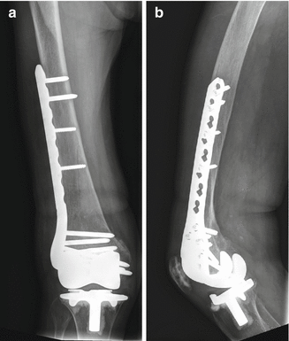
Anteroposterior (a) and lateral (b) radiographs of a supracondylar femoral fracture (Rorabeck [27] type II) (Courtesy of Mr. Nadeem Mushtaq, St Mary’s Hospital, London)
19.3.1 Periprosthetic Femoral Fractures
The femur is the commonest site of periprosthetic fracture and is the most studied. Several classification systems exist, based either on the configuration of the fracture, the involvement of the prosthesis or a combination of the two. Two descriptive classification systems, by Di Giola and Rubash and Chen et al., classify fractures based on the degree of displacement and comminution; neither provides information about the stability of the implant, and both are of only limited usefulness in terms of management [25, 26].
Lewis and Rorabeck classify periprosthetic femoral fractures into three types: types I and II are non-displaced and displaced fractures, respectively, adjacent to a well-fixed prosthesis; type III is any fracture adjacent to a loose prosthesis [27]. The authors suggest that type I fractures be treated nonoperatively, type II fractures be treated with fixation and type III fractures be treated with revision surgery. A similar system is proposed by Kim et al.: type I fractures occur adjacent to well-fixed implants in good quality bone; the authors suggest either conservative management (in reducible fractures, which they term Ia) or fixation (in irreducible, type Ib fractures) [28]. Type II and III fractures involve loose implants, either with good quality bone (type II, which can be revised using a long-stem component) or with poor bone (type III, where an endoprosthesis is required). Su et al. describe a classification system based on fracture position: type I is proximal to the femoral component, type II is at the proximal part of the component, and type III is distal to the proximal part of the component [29]. They suggest that type I fractures should be treated with an intramedullary nail, type II with a fixed-angle plate and type III with revision arthroplasty.
19.3.2 Periprosthetic Tibial Fractures
Fractures of the tibia are less common than fractures in the femur but are more likely to occur intraoperatively [11]. The classification system of Felix et al. works on similar principles to the main classification systems for femoral fractures, classifying fractures by their position and the degree of involvement of the prosthesis [13]. Type I fractures are splits or depressions in the tibial plateau; type II fractures are adjacent to the tibial stem; and type III fractures are distal to the stem of the prosthesis. Fractures of the tibial tuberosity are termed type IV. In each case, the prosthesis can be classed as stable (A) or unstable (B); as with the femur, fractures adjacent to stable prostheses are best treated with fixation whilst if the implant is loose, revision is recommended. Fractures occurring during implantation are given the suffix C; in these cases, on-table revision to a stemmed prosthesis is recommended.
19.3.3 Periprosthetic Patellar Fractures
Periprosthetic patellar fractures overwhelmingly occur in patellae which have been resurfaced: a systematic review of 582 cases reported that only five cases (0.9 % of the total) were in unresurfaced patellae [10]. Unlike other fractures about the knee, periprosthetic patellar fractures are often asymptomatic: in the same systematic review, in 539 cases where the mechanism of injury was documented, 476 (88.3 %) were diagnosed during routine follow-up and had no history of trauma [10].
As with the femur, multiple classification systems have been proposed for the patella [30]. The most commonly used system is that of Ortiguera and Berry, which resulted from a study of 85 fractures in the Mayo Clinic database [10, 12]. Types I and II have a stable prosthesis and are classified according to the state of the extensor mechanism. Type I fractures have an intact extensor mechanism and may be managed nonoperatively. In type II fractures, the extensor mechanism is disrupted, and the authors recommend operative fixation or patellectomy. In type III fractures, the implant is loose. If the bone stock is good (type IIIa), fixation and implant revision can be attempted; if the bone stock is poor (type IIIb), the authors recommend removal of the patellar component and patelloplasty or complete patellectomy.
19.3.4 The Universal Classification System
The Universal Classification System (UCS) is a classification system intended to apply to any periprosthetic fracture in any bone [30]. Similar to the Vancouver classification of periprosthetic fractures about the hip, the UCS classifies fractures into types A–C based on position within the bone, with the addition of types D, E and F (Table 19.1). It is straightforward and intuitive and has the advantage of accounting for fractures (such as inter-prosthetic fractures) not classifiable using the other systems discussed here. Unlike the other classification systems, it has been examined for intra- and interobserver reliability in both experts and trainees [31]. Interobserver reliability was substantial in both groups (Kappa 0.741 and 0.765, respectively), and intraobserver reliability was near perfect (Kappa 0.898 and 0.878). The UCS is in its infancy and has not been used widely in studies of periprosthetic fracture to date; in this chapter, we will use the existing classification systems outlined above.
Type | Description | Management |
|---|---|---|
A | Fracture of apophysis/protuberance | Fix if displaced and functionally important |
B1 | Fracture of implant bed, well-fixed implant | Osteosynthesis |
B2 | Fracture of implant bed, loose implant | Revision with long-stem implant |
B3 | Fracture of implant bed, loose implant with poor bone stock | Complex reconstruction (e.g. tumour prosthesis) |
C | Fracture of bone, distant from implant | Treat as if implant not present |
D | Fracture between two implants | Treat as appropriate for each implant |
E | Fracture of two bones with implants (e.g. tibia and femur in floating knee) | Treat as appropriate for each implant |
F | Fracture of unresurfaced bone adjacent to joint replacement (e.g. fracture of unresurfaced patellar) | Favour conservative management with later resurfacing |
19.4 Management of Periprosthetic Fractures
19.4.1 Periprosthetic Femoral Fractures
Periprosthetic femoral fractures often occur in elderly patients with significant medical co-morbidities, and reconstruction represents a significant surgical insult [3]. The mortality for periprosthetic femoral fractures is similar to that of hip fracture, and a delay in surgical management increases the risk of death following fixation [1, 3]. Nonsurgical treatment of femoral fractures is associated with a higher rate of malunion than surgical treatment [32]. As such, in the femur, nonsurgical treatment should be reserved for undisplaced fractures and those occurring in moribund patients [32].
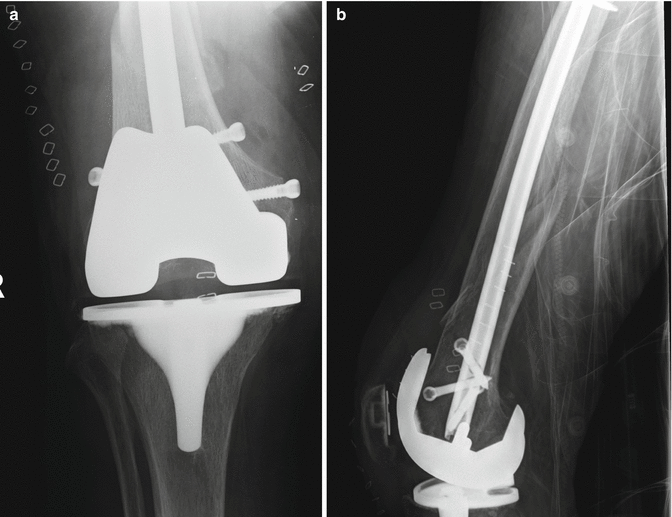
Fixation of periprosthetic femoral fractures in the presence of a stable implant may use locked intramedullary nailing or plate osteosynthesis (Figs. 19.2 and 19.3) [32]. A high complication rate after locking plate fixation has been reported by Ebraheim et al. [2]. Twenty-seven patients with periprosthetic distal femur fractures after TKAs were treated using a distal femoral locking plate. The average time for union and weight bearing was 4.5 months. The union rate was 89 %. Thirty-seven percent experienced complications, with two delayed unions (7.4 %), one non-union (3.7 %) and seven fixation failures (26 %). Cases where the implant is loose necessitate treatment with revision TKA, either stemmed revision implants or megaprostheses, depending on the fracture type and bone stock [7].


Fig. 19.2
Anteroposterior (a) and lateral (b) radiographs of the fracture from Fig. 19.1 following fixation with a lateral locking plate

Fig. 19.3
Anteroposterior (a) and lateral (b) radiographs of a Rorabeck [27] type III femoral fracture following fixation with a retrograde intramedullary nail (Courtesy of Mr. Rajarshi Bhattacharya, St Mary’s Hospital, London)
There are no randomised studies and few prospective comparative studies comparing the outcomes of plate or nail fixation for periprosthetic femoral fractures. Whilst locking plates have the benefit of allowing a more anatomical reduction, there is less soft tissue dissection involved with intramedullary nailing (although this is mitigated in part in minimally invasive plating techniques). Retrograde nailing can only be employed in implants with open intercondylar notches of a sufficient size to take a nail; a compatibility guide has recently been published to aid the identification of such implants [33].
A systematic review of 29 non-comparative case series prior to 2006 (a total of 415 cases) demonstrated significantly higher rates of union and lower rates of further surgery after intramedullary nailing compared to conventional (non-locking) plates [6]. The same study suggested that the results of locking plates are superior to non-locking plates, but the numbers were small, and the difference was nonsignificant.
Eight recent studies compare locking plate to nail fixation. All are small, retrospective studies, and none are randomised, making selection bias amongst the treatment groups likely. Overall, there remains a high rate of failure, and there is little to choose between the two fixation methods. Studies of Gondalia et al. (42 patients), Kilucoglu et al. (16), Hou et al. (40) and Wick et al. (18) report similar outcomes for both modalities [34–37]. Studies of Large et al. (40 patients) and Horneff et al. (63) suggest that locked plating leads to higher rates of union and lower rates of reoperation (although the control group of Large’s study also included fractures treated using non-locked plating) [38, 39]; studies of Meneghini et al. (95 patients) and Aldrian et al. (86) report higher rates of union with intramedullary fixation [40, 41]. Minimally invasive plate fixation may produce higher rates of union compared to open surgery, but the evidence is not strong [42, 43].
Revision surgery can be employed either as a primary management strategy in patients with loose implants or who are unable to tolerate prolonged periods of immobilisation [7] or as a treatment for failed primary fixation [44]. Whilst attempting primary fixation preserves bone stock, the use of revision prostheses in the acute setting reduces the risk of reoperation and is associated with a lower rate of complications compared to revision for failed fixation [45]. There are few studies comparing the two strategies; one retrospective study of 69 patients reports similar survival and functional outcome in patients managed with fixation and revision [46]. However, a series of revision arthroplasty in this setting report high complication rates, and revision should not be undertaken lightly in light of the effectiveness of osteosynthesis in well-fixed prostheses [47, 48].
19.4.2 Periprosthetic Tibial Fractures
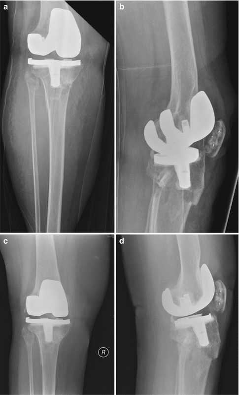
Periprosthetic tibial fractures are substantially less common than femoral factures, and there is much less literature available to guide management. Felix et al. describe an algorithm for the treatment of tibial fractures based on their own classification system (which is described above) [13]. They suggest conservative management in most fractures in the presence of a secure prosthesis and revision surgery when the prosthesis is loose, with only fractures with marked displacement requiring fixation (Fig. 19.4).


Fig. 19.4


Periprosthetic tibial fracture managed nonoperatively: anteroposterior (a) and lateral (b) radiographs at the time of injury. Anteroposterior (c) and lateral (d) radiographs at 3 months with position maintained and evidence of early healing (Courtesy of Mr. Robin Strachan, Charing Cross Hospital, London)
Stay updated, free articles. Join our Telegram channel

Full access? Get Clinical Tree


