Peripheral Tear of the Triangular Fibrocartilage Complex from the Ulna: Arthroscopic Repair

17 Peripheral Tear of the Triangular


Indications


  • Peripheral tear of the triangular fibrocartilage complex (TFCC) from its attachments to the ulna or the joint capsule.

Pitfall


May not restore stability to the distal radioulnar joint (DRUJ) in cases of advanced or chronic instability


Technique


  • The arthroscope is placed in the 3–4 or 4–5 portal depending on the size of the wrist and visualization.
  • A probe is inserted through the 6R or 6U portal. Loss of normal TFCC tension under probe pressure, the so-called trampoline test, is a sign of a peripheral tear.

Pearl


When making portals, nick the skin and spread the subcutaneous tissue to prevent cutaneous nerve and tendon injury.


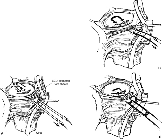
  • A 1.5 cm skin incision is made just dorsal to the ulnar styloid. The extensor retinaculum and extensor carpi ulnaris (ECU) subsheath are opened 1 cm and the tendon retracted enough to visualize the floor of the sixth compartment.

Pitfall


Protect any branches of the dorsal sensory ulnar nerve that pass nearby.


image

Only gold members can continue reading. Log In or Register to continue

Stay updated, free articles. Join our Telegram channel

Mar 21, 2017 | Posted by in MUSCULOSKELETAL MEDICINE | Comments Off on Peripheral Tear of the Triangular Fibrocartilage Complex from the Ulna: Arthroscopic Repair

Full access? Get Clinical Tree

Get Clinical Tree app for offline access