11 Pelvis
Introduction
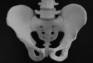
The sacroiliac joint as a source of pain and dysfunction is a subject of controversy.1–6 Many authors implicate the sacroiliac joint as a cause of low back pain,6–19 but there is disagreement as to the exact prevalence of sacroiliac joint pain within the low back pain population. It is estimated that 5–15% of chronic low back pain may involve the sacroiliac joint.14,16,20 Although many practitioners believe the sacroiliac joint is a source of pain and dysfunction and treat perceived sacroiliac lesions, there is no general agreement concerning the different diagnostic tests and their validity in determining somatic dysfunction of the pelvis (Fig. 11.1).16,21–33 Assessment of the sacroiliac joint in the male population may be confounded by joint fusion, which is present in 5.8% of males up to the age of 39 increasing to 46.7% of males over 80 years of age.34
A large number of diagnostic tests exist to evaluate the sacroiliac joint. Motion tests and static palpation of bony landmarks have generally shown poor inter-observer reliability. Pain provocation tests have shown better reliability,35 with clusters of pain provocation tests showing even greater reliability.35–38 Arab et al39 suggest that composites of motion palpation and provocation tests may be useful in clinical practice. Laslett40 suggests that the combination of non-centralization of pain on repeated trunk movements with three or more sacroiliac joint provocation tests that reproduce the patient’s familiar pain might help to differentiate sacroiliac joint pain from other painful conditions.
Various models of sacroiliac motion have been proposed and there have been a number of studies relating to mobility in the sacroiliac joint,41–48 but the precise nature of normal motion remains unclear.1,4,17,49,50 There is significant variation in sacroiliac joint movement both between individuals and within individuals when mobility of one sacroiliac joint is compared with the other side.43 Mobility alters with age and can increase during pregnancy.
At our present state of knowledge, what model should guide our clinical decision making to incorporate high-velocity low-amplitude (HVLA) thrust techniques within a treatment regimen for somatic dysfunction of the pelvis? There are a number of different biomechanical models used to determine the nature of any pelvic dysfunction.51–55 These vary from very complex to less complex with no research evidence as to their clinical utility. Greenman54 describes a number of possible pelvic girdle dysfunctions, which are listed in Box 11.1.
A number of manual medicine texts56–61 refer to the use of HVLA thrust techniques to the joints of the pelvis, but there is little evidence that cavitation is uniformly associated with these procedures. When an audible release does occur, its site of origin remains open to speculation. Studies undertaken to measure the effects of manipulation upon the sacroiliac joints provide contradictory findings. Roentgen stereophotogrammetric analysis was unable to detect altered position of the sacroiliac joint post manipulation despite normalization of different types of clinical tests.62 However, an alteration in pelvic tilt was identified post manipulation in one study of patients with low back pain63 and a further study demonstrated an immediate improvement in iliac crest symmetry immediately after manipulation.64 A review of the literature did not reveal any randomized controlled trials investigating the use of HVLA thrust techniques for pelvic girdle pain.
Somatic dysfunction is identified by the S-T-A-R-T of diagnosis:
Stay updated, free articles. Join our Telegram channel

Full access? Get Clinical Tree


