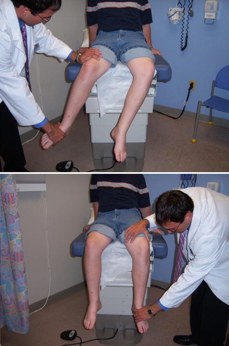
Fig. 12.1
Child with SCFE. Note the increased external rotation of the hip when the leg is hanging

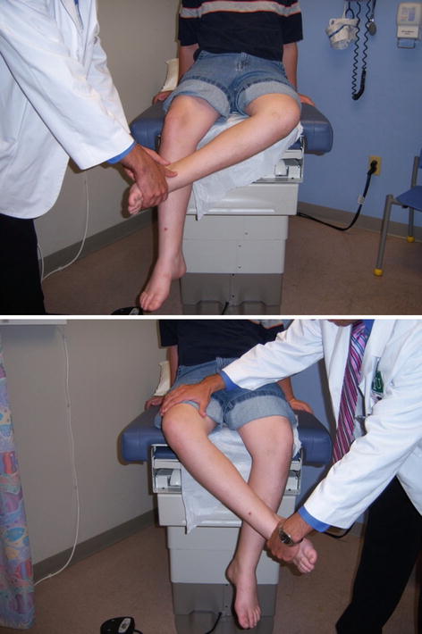
Fig. 12.2
Child with SCFE. Note the decreased internal rotation on testing of the left leg

Fig. 12.3
Child with SCFE. Note the increased external rotation of the left leg compared to the right
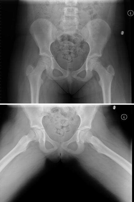
With suspected SCFE, immediate X-rays are necessary. An AP pelvis X-ray (visualizes both hips) and frog view should be obtained as long as the patient can walk (see Fig. 12.4). Those who cannot walk can have bilateral cross-table lateral views instead. X-rays should be read immediately, and if SCFE is diagnosed or suspected, the patient should be made non-weight bearing and an immediate consultation with an orthopedic surgeon should be made. Surgery is usually performed within 24 h. For cases with equivocal X-ray results, phone consultation with the orthopedist is recommended as it is possible to catch the disorder in the “pre-slip” phase. With surgical intervention, outcomes are excellent. Delayed treatment can lead to hip deformity or avascular necrosis of the femoral head, necessitating multiple surgeries and early arthroplasty.


Fig. 12.4
X-ray findings in SCFE. Note that the SCFE is not obvious in AP view; lateral or frogleg view is required to see the anomaly
Bowlegs and Knock-Knees
Bowlegs (genu varum) and knock-knees (genu valgum) are common reasons for parents to request evaluation of their children. The vast majority of these children do not require any intervention, as their condition simply represents normal childhood growth and development. Babies are generally born with mild bowing of the tibia that becomes more noticeable when they begin walking and then usually resolves around 2 years of age. The legs then begin to have a knock-knee appearance, which usually peaks around age 3–4 and resolves by age 6–8.
Genu Varum (Bowlegs)
In toddlers 2 years of age and younger with normal height and weight, and who have no red flags, the bowing is likely physiologic and should resolve spontaneously. If red flag conditions are present (Table 12.1), referral to a pediatric orthopedist should be made. Conditions such as rickets, skeletal dysplasia, Blount’s disease, and others must be considered.
Table 12.1
Red flags for bowlegs
Age >2 years |
Decreased calcium and vitamin D intake (dietary) or lack of sun exposure |
Family history of short stature, adult bowlegs, severe kidney disease, or neurofibromatosis |
Child with short stature, underweight, or significant overweight |
Severe tibia deformity (look for dimple on the apex of the bow) and/or small, deformed foot |
Unilateral deformity or significant asymmetry |
“Thrust” during gait (knee looks like it’s buckling) |
Genu Valgum (Knock-Knees)
Children aged 2–6 who are of normal height and weight, who are otherwise healthy, and who have no red flags most likely have physiologic knock-knees, which should resolve spontaneously. If red flags are present (Table 12.2), referral to a pediatric orthopedist should be made.
Table 12.2


Red flags for knock-knees
Stay updated, free articles. Join our Telegram channel

Full access? Get Clinical Tree


