, Paul D. Siney1 and Patricia A. Fleming1
(1)
The John Charnley Research Institute Wrightington Hospital, Wigan, Lancashire, UK
Use of radio-opaque cement and availability of good quality serial radiographs, showing all of the stem and cement, is essential in order to study the patterns of failure of stem fixation. The three basic patterns can be recognised: slip, tilt and pivot. Although obvious, when typical, an overlap between the types is common.
Slip
Stem Within the Cement Mantle
Limited taper slip of the stem within the cement mantle is the principle behind the design of the C-Stem and the surgical technique of stem fixation. The slip can be recognised, radiologically, by the separation of the stem from the cement proximally on the lateral aspect (Fig. 21.1).
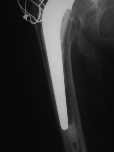
Fig. 21.1
Radiograph showing a slip of the femoral stem within cement mantle. Separation of stem from cement denotes stem slip or/and stem tilt into varus
If the stem was supported distally by cement then fracture of the cement at the tip of the stem will be seen [1] (Fig. 21.2).
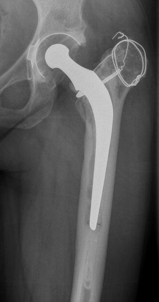
Fig. 21.2
Radiograph showing fracture cement at the tip of the femoral stem, separation of stem from cement and endosteal cavitation medially
These changes may be observed early, usually within the first post-operative year. They may be innocuous. The problem, however, is the real possibility of fragmentation of the cement in the proximal medial region. Regular monitoring is essential. Such changes may herald endosteal cavitation, stem loosening or fracture.
Related posts:


Stay updated, free articles. Join our Telegram channel

Full access? Get Clinical Tree


