, Paul D. Siney1 and Patricia A. Fleming1
(1)
The John Charnley Research Institute Wrightington Hospital, Wigan, Lancashire, UK
Consistency of materials, design and the surgical technique of the Charnley LFA and the long term follow-up results offer detailed information which can be used to set practical guidelines for follow-up.
The clinical results have been recorded since the beginning of the operation in November 1962 up to the end of December 2014, where 25,753 primary LFAs had been carried out. Over this 52 year period, 1,433 (5.6 %) hips had been revised. Survivorship analysis [1] to 37 years where a minimum of 40 hips [2] were still attending was calculated.
The Reason for and the Frequency of Follow-Up
The reasons for follow-up with serial radiographs is to identify problems early and offer revision surgery before complex mechanical failures present clinically. A balance must be struck between the frequency of visits and the value of information obtained. It is in this context that the knowledge of patterns of failure, both by the specific mode and the particular design and technique, is essential. An immediate post-operative radiograph (a-p and lateral) is mandatory. A record, at 1 year after the operation, is an invaluable baseline for comparisons. No specific time intervals can be recommended and “tailoring” the pattern for individual patients becomes the practice. This should be based on the knowledge of underlying pathology, radiographic appearances and progress within the first year after surgery, patient’s activity level and the rate of wear of the UHMWPE cup. It is for these reasons that continuity of observer method is essential. Intervals longer than 2 years proved unsatisfactory. Logistics of planning more than 2 years ahead were administratively too complex and non-attendance levels too high and thus a waste of resources with the increasing access to the internet a timely reminder to attend follow-up may be of help. Free access, at short notice, however, must always be available.
Anticipating Problems
Pain relief, after total hip arthroplasty, is so spectacular that if the patient’s symptoms have not been relieved three possibilities must be considered:
Complications of surgery.
Incorrect identification of the original pathology
Inappropriate patient selection
Complications at Surgery
Attention must be focused on identifying or excluding complications. It is in this context that the availability of good quality sequential radiographs is essential.
Deep Infection
Patients at risk for deep infection include those with previous hip operations, diabetics, patients with rheumatoid arthritis, psoriasis. History of post-surgery urinary tract infection, catheterisation – and in the male – prostatectomy.
Post-operative pain and delayed wound healing is the most sinister combination.
Radiologically – trochanteric separation, wedge erosion of the bone-cement junction at the medial femoral neck, early progressive demarcation of the cup, periostitis at the level of the stem tip and endosteal cavitation at the same level. With any of these, either singly or in combination, infection must be considered until proved otherwise (Fig. 42.1).
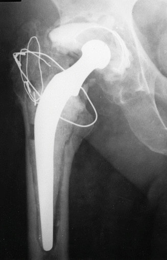
Fig. 42.1
Radiograph of an infected LFA
Clinical experience indicates that a diagnosis of deep infection is usually suspected, even if not established, within 1 year in the majority of cases with 80.6 % of deep infections having been revised by the eighth year post-op (Fig. 42.2). It is this group of patients that demands regular review – with radiographs and relevant blood tests – every 3 months at least. Although given time infection will declare itself, a high index of suspicion must remain if progress after surgery is slow or symptoms persist. Survivorship at 37 years follow-up was 92.56 % (Fig. 42.2).
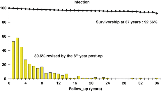
Fig. 42.2
Revision for infection. Incidence and survivorship analysis
Dislocation
The period at risk is days or weeks after surgery. The complication is usually “self-reporting” if associated with pain and loss of mobility and always if manipulation was required after confirming the diagnosis by a radiograph. Episodes of subluxation, or even dislocation, with spontaneous relocation, may be missed. It is for those reasons that this complication remains under-reported. Detailed history of the activity leading to the problem is very helpful. Soft tissue trauma due to tearing of the capsule by the subluxed, dislocated head, results in pain which may persist for days or even weeks. Review within 6 weeks or so is recommended to either confirm the diagnosis, establish recurrent dislocation as the problem or exclude the more sinister diagnosis – deep infection. Over half of the revisions for dislocation were within 6 years of surgery with revision after the eighth year rare, and often the consequence of wear and resultant cup loosening. The survivorship at 37 years follow-up was 97.34 % (Fig. 42.3).
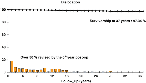
Fig. 42.3
Revision for dislocation. Incidence and survivorship
Fracture of the Stem
Fracture of the stem, was at one stage, the most common indication for revision of the Charnley LFA. The first cases presented in 1968 – 6 years after the introduction of the operation into routine clinical practice. At this stage some 2,500 hips had been implanted. The period at risk was 4–13 years after surgery with survivorship at 37 years – 93.33 % (Fig. 42.4). Radiographic “at risk” signs may be present earlier: separation of the stem from the cement – proximally-laterally, with poor cement mantle proximally-medially. Slip of the stem within the cement mantle, with failure of the cement at the tip of the stem, may lead to fragmentation of the cement – proximally medially.
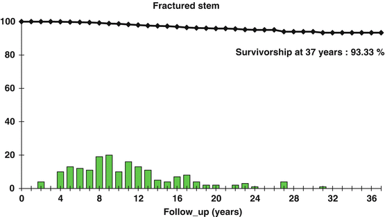
Fig. 42.4
Revision for fractured stem. Incidence and survivorship
Lack of clearance at the anatomical calcar, and hence absence of the stem support proximally posteriorly, is the common finding. Mechanically – lack or loss of the proximal stem support in the presence of good distal fixation is the typical picture. Clinically – active, heavy male after years of excellent function presents with sudden pain which may come on after an apparently normal act like stepping down a step or a sudden turn (Fig. 42.5).
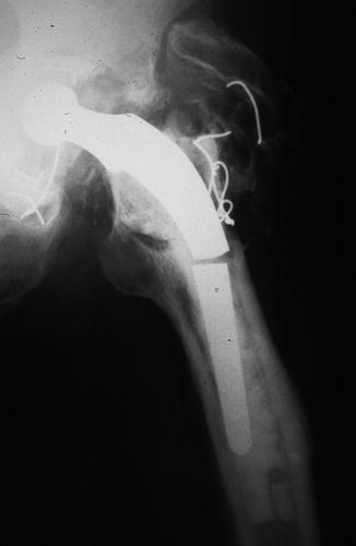
Fig. 42.5
Radiograph showing stem fracture
This signals the end point of what is fatigue fracture – the mechanism is bending-torsion of the proximal part of the stem with a well fixed distal portion [3]. The initial pain may settle and divert the attention from the real pathology. Urgent radiograph (AP and lateral) is essential because the short free stem fragment may lead to fracture of the proximal femur. The mechanism of stem fracture is basically the same as resulting in slipped upper femoral epiphysis, fracture neck of the femur, localisation of Legg Calve Perthes and avascular necrosis lesions, as well as some cases of osteoarthritis.
Fractured stem after primary LFA is now largely of historical interest. Improved materials (ORTRON: DePuy International, Leeds UK), stem design [4], and surgical technique [5] have eliminated the problem in primary surgery. The effects of proximal strain shielding of the femur present later as a more serious complication of periprosthetic fracture of the femur.
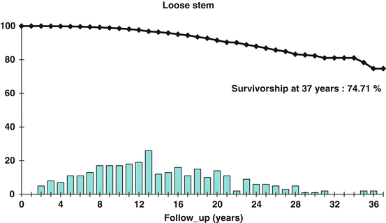
Loose Stem
Loosening of the cemented stem is unusual within the first 4 years and may be largely considered to be due to inadequate fixation or alternatively excessive loading in heavy patients. The “at risk” radiographic appearances have been documented: [6] demarcation of cement at the tip of the stem, fracture cement at the tip of the stem, valgus stem position. Separation of the stem from cement – not significant as a single factor, gains significance when in combination with other signs described. Endosteal cavitation is rarely seen in the first year. Its appearance, at any stage, spells failure and frequent reviews are essential. Stems that are revised for loosening fail between the 5th and 20th year with a survivorship at 37 years follow-up of 74.71 % (Fig. 42.6).