Patient-Specific Unicompartmental and Bicompartmental Resurfacing Arthroplasty

Chapter 16 Patient-Specific Unicompartmental and Bicompartmental Resurfacing Arthroplasty


Introduction


Total knee arthroplasty has become one of the most common surgical procedures due to its clinical success and longevity. In the United States, more than 500,000 primary total knee replacements were performed in 2006.1 This volume is expected to grow to 3.48 million by 2030.2


In the early 2000s, the average age of patients undergoing knee replacements was between 67 and 68 years. Numerous studies and anecdotal experience from surgeons have reported a rise in the incidence of primary osteoarthritis (OA) in younger patients. Factors such as an aging baby boomer population with more active lifestyles, the rise of sports-related injuries in younger populations as a precursor to osteoarthritis, and a more demanding patient base unwilling to modify their activity levels appear to be potential causes of the increase in younger patients seen by surgeons.


In younger patients with moderate OA, a different set of considerations enters into the calculation for the appropriate treatment approach. Early intervention patients often are more active, have a higher potential for revision later in life, and exhibit patterns of OA that may be more localized. The ability to provide a joint-preserving option that can bridge the patient to a traditional total knee replacement is appealing.


In this chapter, I describe my experience with an imaging-based approach to partial knee resurfacing as a treatment for early intervention patients. I briefly describe the technology and then the surgical technique using patient-specific custom instruments.


Imaging technology


The process for building patient-specific implants and instruments begins with a preoperative computed tomographic (CT) scan performed on commonly available machines (Figure 16–1). The scan, which includes partial views of the hip and ankle, is converted into a virtual three-dimensional model of the patient’s knee. The partial views of the hip and ankle are used to align the implant and cut guides in relation to the patient’s anatomic and mechanical axes.
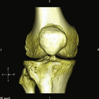


Design engineers at the manufacturer ConforMIS (Burlington, MA) use computer-aided design software to perform virtual osteophyte removal and to identify key anatomic landmarks. The proprietary software then generates a matching surface topography for the femur from the scan data and an outline of the tibia at the expected resection level.


The surface topography is used as the undersurface of the femoral implant, allowing the component to rest on the subchondral bone with close conformity to the patient’s native anatomy. The articulating surface follows the anatomy but with an engineered curve in the coronal plane to minimize contact stress with the tibial surface. The femoral implant thickness is 3.5 mm, representing the native cartilage that is lost. Therefore, it is a resurfacing implant and exactly restores the joint line. The tibial implant is designed to match the outline of the cortical shell on the resected tibia. The custom nature of the implant design allows for complete cortical rim coverage in each patient. The tibial thickness is 9 mm: 7 mm polyethylene and 2 mm metal base plate.


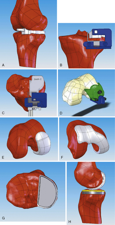
One of the more unusual aspects of surgery using patient-specific implants is the instrumentation set, which consists of a small number of guides that are generated for one-time use with each implant. The cut guides are matched to the patient’s anatomy using the same process as the implants, thus eliminating intraoperative sizing and allowing for tactile feedback to help establish positioning. The instruments are prenavigated using the scan data so that all cuts and drill holes are set with respect to the axis (Figure 16–2).


The design algorithms have been developed for a unicompartmental and a bicompartmental option. The unicompartmental option is well known in orthopedics, but the bicompartmental option is relatively novel. It allows for treatment of the patellofemoral joint in addition to one of the tibiofemoral compartments, and it provides a different option for treating OA in younger patients. Both implants can be designed for either the medial or the lateral compartment. Because the prosthetic trochlea surface is precisely that of the patient, the patella can be left unresurfaced and will have good congruence to the trochlea.


These implants and instruments are manufactured using processes that are designed to be cost competitive at single-unit or low-volume production. The implants are made of standard orthopedic materials, such as cobalt-chromium-molybdenum alloy and ultra-high-molecular-weight polyethylene (UHMWPE), but the cutting guides are made of disposable, engineered materials such as plastic or nylon. The production process takes 4 to 6 weeks for completion.


Surgical technique


The surgical technique described here uses a medial unicompartmental replacement to illustrate the differences of a custom implant with custom disposable jigs. The bicompartmental implant uses a surgical technique very similar to that for the unicompartmental implant except for patellofemoral preparation and fixation.


Jun 19, 2016 | Posted by in MUSCULOSKELETAL MEDICINE | Comments Off on Patient-Specific Unicompartmental and Bicompartmental Resurfacing Arthroplasty

Full access? Get Clinical Tree

Get Clinical Tree app for offline access