Fig. 13.1
Instrument (pink shape) molded on the bone model using acrylic. The instrument presents a flat surface indicating the target osteotomy
Joint Arthroplasties
Several joint arthroplasties are now benefitting from PSI to be planned and positioned. Several research projects have led to commercial products. For the shoulder arthroplasty, Imascap (Brest, France) has developed a software to give the surgeon the ability to perform its virtual pre-operative planning. This software is now proposed by Tornier (Montbonnot Saint-Martin, France) as the Blueprint® solution. Imascap has also developed and brought onto the market a PSI to transfer the planning into the operative room.
Hip prosthesis is also benefitting from PSI to increase accuracy of implant positioning. Several research projects are validating the concept [20, 34, 43]. Increased accuracy has been proven when using the PSI, showing that the technology may be useful in a clinical situation. However, there is no commercial application to date even if a patent has been registered in 2013 and a clinical study launched in the next few months.
The situation is more advanced for ankle arthroplasty were developments have been validated and transferred to a commercial product. Wright Medical has put on the market the Prophecy® Inbone® to position their prosthesis. Berlet et al. [3] have observed a high repeatability in positioning the instrument, leading to a final implant positioning within ± 3° when compared to the planned position. However, the solution is not an actual positioning instrument and clinical data are not available yet.
Bone Tumor Surgery
In tumor surgery, surgical excision must be highly accurate to ensure the total removal of the pathologic tissue without infraction of the tumor. Other way, a local recurrence of the tumor can arise leading to a failure of the initial treatment. The conventional method has shown unsufficient clinical results with a local recurrence rate observed in 28–35 % in case of pelvic tumors [9]. These clinical results have been confirmed by in vitro experiments leading to intralesional resections on plastic pelves [5, 6]. Preoperative assistances have been developed to plan the tumor removal. Tumor extension is delineated on the MRI and merged with the CTscan to combine anatomical and functional data. Based on the generated 3D view, cutting trajectories are chosen including a user-defined safe Margin (Fig. 13.2) [26]. When needed, a reconstruction strategy can be planned as well, using either a frozen allograft [27] or a commercial implant. The transfer into the operating theater was made possible by using a customized optical navigation system. The overall accuracy of the process has been assessed on plastic pelves [5]. A significant improvement has been shown with a reduced error during bone cuttings from 11.2 mm down to 2.8 mm (p < 0.001). The system has been used to surgically treat a small number of patients [14]. The technique has been more widely described by Docquier et al. the creators of the overall system [13].
Fig. 13.2
Preoperative planning of a tumor surgery. (a) Shows the cutting trajectories around the tumor, including a safe margin. (b) Shows the instrument designed to resect the tumor. Allograft selection can be performed virtually (c) according to reconstruction needs. An instrument can also be designed to actually cut the allograft (d)
The previously described resurgence of PSI has conducted the developers of the assistances to move toward this new accessible technique. PSI have been tested on plastic pelves to assess the feasibility of tumor resection and estimate their accuracy [7]. The mean accuracy was below 2 mm, showing a significant improvement when compared to the conventional method and a non-significant improvement regarding the optical navigation. Since then, PSI have been used on actual patients to treat several bone (Figs. 13.3 and 13.4). Their excellent accuracy has permitted to decrease the target safe margin from 10 mm (standard desired safe margin) down to 4 mm in some cases. The objective of decreasing the target safe margin is crucial since it allows the preservation of important anatomical structures such as joints, ligament insertions or nerves.
Fig. 13.3
Bone models and instruments manufactured. Bone models from the patient (a) and from the allograft (c) can be manufactured and sterilized. Instruments positioning are checked before the surgery to ensure a satisfying use (b, d)
Fig. 13.4
Instruments use during the surgery. The instrument for resection is positioned onto the bone and fix using KWires (a). The process is repeated for the allograft (b). After allograft cutting, the accuracy is checked on the allograft model (c). Finally, the cut allograft is impacted in the defect (d). A perfect fit is obtained with contact for each of the 7 cutting planes
A spin-off company from the Université catholique de Louvain (3D-Side, Belgium) has been launched to put the technology on the market. To date, 55 patients have been successfully treated in several European countries. Clinical series have been reported in the literature showing good oncological results [2, 16]. No local recurrence linked to a bone contamination has been observed postoperatively even if the post-operative follow up is too short to draw strong conclusions. A local recurrence has arose because of a contaminated soft tissue margin. R0 safe margins have been systematically obtained except in one case where the tumor has been morselized to urgently extract it from the patient who was suffering from severe bleeding and poor cardiovascular conditions. A further cost-efficiency study will be led to assess a potential financial benefit of the technique regarding the prevented cost of local recurrence.
Corrective Osteotomies
Patients who have suffered from a bone fracture usually recover a fully normal limb function. In some cases, a non-anatomical bone fusion can be observed yielding to a limited function of the involved limb. When the limitation prevents a normal everyday life, a corrective surgery is indicated. The required correction is often a complex biplanar bone cutting representing a solid angle. By using a conventional manual method, the obtained correction is often suboptimal, leading to an over- or a sub-correction. A 3D simulation of the cuttings is highly helpful to visualize the initial position and estimate the appropriate correction that should be brought.
Some companies (Materialise, 3D-side, Cartis) and academic research projects propose a 3D analysis showing a reconstruction of the bone and proposing a strategy to restore a normal anatomy [11, 12, 41]. The retained surgical option is transferred into the operating room by using PSI. Three different approaches can be adopted.
The first method is based on a PSI that serves as hole driller and saw guide. Firstly, holes are drilled into the involved bone, representing the trajectories of future screws. Secondly, the PSI is used to cut the bone thanks to a slot guiding a saw blade. The PSI is removed from the bone and the reconstruction is performed by inserting a dedicated plate and the pre-drilled screws. This technique requires the use of 3D models from screws and plates of an implant manufacturer during the preoperative planning. Also, the technique does not allow any change in plate and screws sizes.
In the second philosophy, the PSI is positioned onto the bone and fixed in place by using two-by-two parallel k-Wires. The PSI indicate the osteotomy to be performed, guiding the saw blade (Fig. 13.5). After bone cutting, the k-Wires are parallelized to obtain the correct limb alignment as planned preoperatively. This second solution is not dependent from any implant manufacturer and thus any osteosynthesis material can be used to make the osteosynthesis.
Fig. 13.5
Instrument is positionned onto the bone surface (a). KWires are inserted into the cylindric guides to fix them. A flat surface guides the saw blade (b)
Finally, the third method consists in determining a single cut that may correct the anatomy. A PSI allows to perform the cutting and drilling the future screws trajectories. Finally, a patient-specific plate, including a solid angle portion corresponding to the correction that will be brought, is designed to realign the bone segments into an anatomic position. The plate is manufactured by additive manufacturing and implanted during the surgery.
Other Applications
Synostosis
This pathology, abnormal fusion between two bones, usually affects the calcaneonavicular or the talocalcaneal junctions. It occurs in approximatively 1 % of the population, specifically in young people. The treatment is a surgical resection in the coalition zone, removing sufficient portion of bones. The recurrence rate is relatively high because of unsufficient resection. In case of breakage of the healthy joint, hidden by the bones, the foot can be painful. Accuracy can be improved by performing a simulation of the resection and actually create it using a PSI (Fig. 13.6). The planning permits to ensure a complete resection of the degenerative joint. The depth control preventing breaching the healthy joint can be achieved if the saw depth is determined at the stage of preoperative planning. This technique has been reported, as well as early clinical results on 9 patients [10]. No recurrence has been observed at last follow-up.
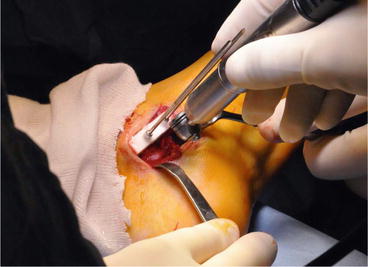
Fig. 13.6
The instrument indicates the osteotomy to perform. The depth control is achieved thanks to a physical stop that prevents the blade from going into the safe joint
Paprovsky Pathology
This pathology is a degenerative process of the hip that can lead to incapacity of walking for the patient. In many cases, the hip cannot be restored properly since bone loss is too extensive to accept a standard hip implant. This surgery was among the first to benefit from PSI. A synthetic metallic implant was designed to fill the hip defect and restore a normal anatomy. The implant is manufactured using additive manufacturing, usually in titanium or Cobalt-chromium alloys. The manufacturing process produces porous surfaces, once in contact with patient’s bone present excellent properties to accept bone ingrowth. Regarding the joint, a finishing is required to produce smooth surfaces allowing a normal function of the joint. An associated instrument is designed to perform an accurate resection and ensure an optimal fitting of the implant with the anatomy. Mobelife, a spin-out from Materialise (Leuven, Belgium) has put this solution on the market in the years 2010. The main disadvantage of the methodology is the high cost of the manufacturing that leads to a very expensive solution for the surgeon. The medical benefit should be balanced against the economic profit that may incur. In some countries, the social system has accepted a reimbursement.
Ilio-Sacral Screws
Stabilization of joint dislocation or sacral fractures is usually performed by inserting screws through the sacro-iliac joint. The accuracy is crucial in this surgery to ensure a bone insertion without breaching sacral nerves. Recently, PSI have been clinically used in 16 patients [8]. Reported results were promising when compared with conventional fluoroscopic insertion. A significant improvement in accuracy has been shown. PSI are particularly suitable for this application since the surfaces of the iliac crest are highly discriminant allowing an easy positioning and an immediate stability of PSI.
Conclusion
Patient-Specific Instruments has deeply modified the orthopedic surgery, bringing new possibilities [32]. Supported by what is considered as the third industrial revolution, namely additive manufacturing, PSI have invaded the operative room. Emphasize must be put on the pre-operative planning which takes a crucial role. Firstly, it has to be as accurate as possible since the assistance is meant to replicate the planning on the actual bone. Secondly, the planning must first conform to the situation that will be met in the operative room. It implies that the surgeon anticipates how surgery will be performed several days or weeks before the actual surgery. For example, the surgical approach must be firmly defined since it has a strong impact on the instrument design or the contact surface with bone. Finally the planning must be performed using 3D data to define cutting or drilling trajectories in the 3D space. This new approach generates new tasks to perform and new tools to understand for the surgeon. That’s why an engineer is often responsible for handling the computerized tools that produces the pre-operative planning. The cooperation between the surgeon and the engineer is thus critical to generate a planning that reaches the desired target and meets the medical requirements.
Related posts:
 Virtual Preoperative Planning
 Computational Image-Guided Technologies in Cranio-Maxillofacial Soft Tissue Planning and Simulation
 Virtual Cranio-Maxillofacial Surgery Planning with Stereo Graphics and Haptics
 Spinal Loading System: A Novel Technique for Assessing Spinal Flexibility in Adolescent Idiopathic Scoliosis
 Navigation in Spinal Surgery
 3D Projection-Based Navigation
Stay updated, free articles. Join our Telegram channel

Full access? Get Clinical Tree


