M. Bradford Henley
Sterile Instruments/Equipment
- Large and small pointed bone reduction clamps (Weber clamps)
- Specialized patellar bone clamps
- Implants
- Cannulated 3.5- or 4.0-mm screws
- 1.0-mm cable or 18-gauge wire
- Mini-fragment screws for free fragments
- Mini-fragment plates (2.0/2.4 mm) for associated coronal plane fracture lines
- Strong nonabsorbable suture with good handling characteristics, such as no. 2 Fiberwire, Ticron or Tevdek.
- Cannulated 3.5- or 4.0-mm screws
- K-wires and wire driver/drill
- Beath pins or Hewson wire passer
- Sterile, removable bump for alternate placement behind knee/heel to obtain knee flexion/full knee extension
Patient Positioning
- Supine on a radiolucent cantilever table.
- Padded ramp under affected extremity to facilitate lateral imaging.
- Bump placed under the ipsilateral hip to limit external rotation of extremity.
- Padded tourniquet placed on the thigh if desired.
Surgical Approach
- Midline longitudinal incision to deep fascia.
- A horizontal “smile” incision may be used for improved cosmesis in simple fracture patterns.
- The medial and lateral ends of the transverse incision should be slightly curved proximally.
- A horizontal “smile” incision may be used for improved cosmesis in simple fracture patterns.
- Fracture is identified and cleansed of clot and fracture debris.
- Flex the knee over the sterile bump to identify and document associated intra-articular pathology, such as chondral injury to the trochlea or femoral condyle.
- Work through lacerations in medial and lateral retinaculae to view and/or palpate articular reduction.
- These can be extended, if needed.
Reduction and Implant Techniques
- Modified tension band.
- Reduction often facilitated with knee in full extension, to relax extensor mechanism especially when there has been retraction of fracture fragments/extensor mechanism.
- Place a sterile bump behind the heel/distal leg.
- Grasp major fragments with small pointed reduction forceps for direct manipulation while an assistant clamps major fragments into place with large pointed reduction clamps or a patellar clamp.
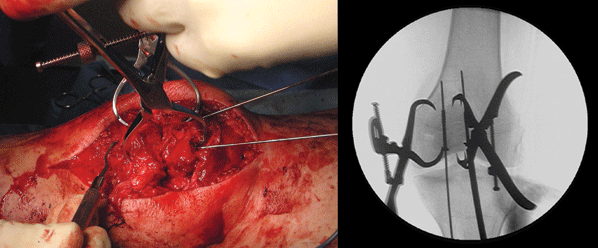
- Specially designed clamps are available with dual prongs on each tine to grasp the bone though quadriceps and patellar tendons (Fig. 18-1).
- Reduction often facilitated with knee in full extension, to relax extensor mechanism especially when there has been retraction of fracture fragments/extensor mechanism.

Figure 18-1. Specialized clamps with two prongs on each tine (large Weber clamp is to the left [medial] and patellar clamp is to the right [lateral] on the C-arm view) facilitate grasping the edge of the patella through tendon(s) for manipulation and stabilization.
![]()
- Fine-tune reduction with dental picks.
- Place multiple K-wires in patella for provisional fixation.
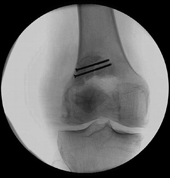
- Lag screws may be placed through additional fragments to reconstruct the patella so as to convert a multifragmentary fracture into a simpler two part fracture with two remaining large fracture fragments and a transverse or short oblique fracture line (Fig. 18-2).

Figure 18-2. Screws placed perpendicular to the fracture lines of additional fragments should be used to sequentially reconstruct the patella into a main proximal and distal fragment.
- A surgical tactic should account for the location of screws in the patella (e.g., screws placed horizontally are placed more anteriorly while longitudinally oriented fixation will be placed closer to the articular surface).
- This will avoid the frustration of screw collisions/deflections that compromise fixation and/or quality of reduction.
- Horizontally placed screws are best placed from lateral to medial as the lateral patellar facet(s) are narrower than the medial facet(s).

Stay updated, free articles. Join our Telegram channel
- This will avoid the frustration of screw collisions/deflections that compromise fixation and/or quality of reduction.

Full access? Get Clinical Tree


