Figure 8.1
Proximal tibia physis anatomy
8.4.2 Diaphyseal Physis
Ossifies at 7 weeks of gestation and extends proximally and distally to meet the other tibial physes.
8.4.3 Distal Tibial Physis
Growth contribution – 5 mm/year.
Closure of the physis follows a predictable pattern. From central to medial, then progresses laterally. The anterolateral area closes last at around 14 years of age. See diagram below (Fig. 8.2).
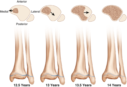
Figure 8.2
Distal tibia physis anatomy
8.5 Blood Supply
The thick periosteum provides a significant proportion of the blood supply to the tibia.
Proximal tibia – this is supplied by a rich extraosseous supply from the popliteal artery, anterior tibial artery (ATA) laterally, and the posterior tibial artery (PTA) medially.
Diaphysis – this is supplied by the nutrient artery, and so is a considerably hypovascular region, particularly posteriorly.
Distal tibia – supplied by the medial ATA and PTA anastomosis.
8.6 Diagnosis
The child will typically present with a history of trauma such as a fall or twisting injury. The mechanism of injury dictates the amount of energy transferred to the tibia and will determine the extent of the bony injury as well as the soft tissue damage. Take note of the child’s age and mobility status. Document whether the child is able to sit independently, crawl, bottom shuffle, cruise or is able to walk. Where the injury sustained is incongruent with history from the parents, non-accidental injury should be considered. Enquire about previous admissions to hospital and carefully document the clinical findings. It is important to liaise with the paediatric team in such cases.
The child will present with a limp, or a complete inability to weight bear on the affected side. Intra-articular injuries may present with a red, hot swollen joint, representing a haemarthrosis caused by ligamentous or bony injury.
8.7 When Examining the Tibia
Inspect for lacerations, haematomas and ecchymosis.
Examine for tenderness, and crepitus.
Examine the knee and ankle joint for effusions, and ligamentous stability.
Perform a complete distal neurological examination.
Assess perfusion of the limb – capillary refill time, dorsalis pedis and posterior tibial pulses.
Look for signs of compartment syndrome.
8.8 Imaging
Anteroposterior and lateral radiographs of the tibia are usually adequate.
Obtain views of the ankle and knee joints.
Assess for comminution, displacement, translation, shortening and angulation.
Computed tomography (CT) should be reserved for complex intra-articular injuries only, as there is a significant radiation dose delivered to the child.
Magnetic resonance imaging (MRI) is used to assess ligamentous and meniscal pathology which are often associated with intra-articular fractures.
8.9 Proximal Tibial Fractures
Injuries affecting the proximal tibia include eminence (avulsion) injuries and tubercle fractures. These are considered in chapter (REFER TO KNEE CHAPTER).
8.9.1 Proximal Metaphysis Fracture (Cozen’s Fracture)
Proximal tibial metaphyseal fractures are caused by a force applied to the lateral aspect of an extended knee, which causes the medial tibial physis to fail in tension. The fibula generally remains intact although it may undergo plastic deformation. The peak incidence of this injury is between 3 and 6 years when the femoral-tibial angle is growing towards valgus. Cozen described the development of a late tibial valgus deformity at around 12–24 months post injury. The aetiology of the deformity in uncertain however several theories have been described:
Mechanical – avulsion of pes anserinus causing loss of medial tethering with intact fibula tethering, interposition of soft tissues within medial fracture gap increasing medial physeal growth, and expanding medial callus formation.
Vascular – increase in blood flow to the medial tibial physis causes increased medial growth (Fig. 8.3).
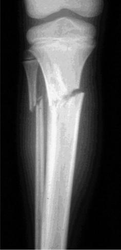
Figure 8.3
Cozen’s fracture
There is no classification system for proximal tibial metaphyseal fractures. They are generally divided into complete or incomplete, displaced or undisplaced.
8.9.1.1 Treatment and Indications for Surgery
Non-operative.
Above knee cast – knee in 5–10° of flexion with varus moulding. Displaced fractures require closed reduction under anaesthesia and application of a varus moulded cast. Immobilise for 6 weeks, but the patient can weight bear after 3 weeks. Wedging of the cast can be used to increase varus force.
Operative.
Interposed soft tissues (pes anserinus, periosteum) within the fracture site can prevent closed reduction. Removal of the soft tissues allows reduction and internal fixation is often not necessary.Related posts:

Stay updated, free articles. Join our Telegram channel

Full access? Get Clinical Tree


