Intrinsic risk factors
Growth-related factors
Susceptibility of growth cartilage to repetitive stress
Adolescent growth spurt
Previous injury
Previous level of conditioning
Anatomic factors
Menstrual dysfunction
Psychological and developmental factors—athlete specific
Extrinsic risk factors
Training workload (rate, intensity, and progression)
Training and competition schedules
Equipment/footwear
Environment
Sport technique
Psychological factors—adult and peer influences
Of all overuse injury risk factors, a previous injury is the strongest predictor [13–22]. This may indicate that a prior injury did not receive complete and comprehensive treatment, creating a susceptibility to repeat injury at the same site. Alternatively, such a situation could alter force dissipation, leading to injury at another site along the kinetic chain.
Characteristics unique to skeletally immature athletes also play a role. Growth cartilage is more vulnerable to injury than mature bone [23–27], particularly during the adolescent growth spurt [27]. In addition, a reduction in size-adjusted bone mineral density that develops before peak height velocity occurs is associated with acute fractures [28]. These factors may also apply to overuse injuries. Injuries such as chronic wrist pain in young gymnasts and proximal humeral physeal stress injury coincide with the expected age range for the adolescent growth spurt [29, 30].
Moreover, asynchronous changes occur during rapid growth that affect the relationships between growth and strength [31]. When combined with the mechanical stresses of training, a milieu distinct to young athletes exists that heightens the risk for overuse injuries.
Amenorrhea is also an established risk factor for overuse injury, specifically BSI [32–37]. The catalyst appears to be inadequate caloric intake leading to a state of hypoestrogenemia resulting in lower bone mineral density, thus lowering the threshold for BSI [38].
Other proposed intrinsic factors such as anatomic alignment, inflexibility, and joint hypermobility have all been cited as risk factors for overuse injury. However, there is little data to substantiate a causal relationship between these factors and injury [39–49].
Among extrinsic factors, training workload has repeatedly been linked to overuse injury.
Both training volume and intensity are significant factors [42, 50–53]. In particular, training more than 16 h per week results in more overuse injuries [51, 54, 55]. A high ratio of workload to rest may also be a specific factor. This can occur during youth sport showcase events and tournaments that include multiple competitive events per day, often over more than 1 day. Studies evaluating this concept are needed [1]. Other extrinsic factors, such as strength and conditioning as well as equipment fit and suitability, may play a role, but lack specific data [1].
Apophysitis
The secondary ossification center where a muscle-tendon unit inserts is known as an apophysis. Apophysitis is an overuse condition causing pain, inflammation, and microtrauma to the apophysis due to traction of the attaching tendon [56]. The skeletally immature athlete is unique in that the apophysis is the weak link in the muscle-tendon-bone unit. There is a lack of data on the actual incidence of apophyseal injuries. These injuries are more common in young athletes participating in running and jumping sports, such as soccer, basketball, and football, especially with use of cleats.
General principles for all apophysitises are described below. Table 7.2 provides a list of common apophyseal injuries.
Table 7.2
Common apophyseal injuries
Apophysis | Eponym | Age (y) | Anatomic site | Exam findings | Treatment specifics (non-avulsion injuries) |
|---|---|---|---|---|---|
Calcaneal | “Sever’s disease” | 8–12 | Calcaneus, Achilles tendon | TTP at calcaneal insertion of the Achilles tendon | Heel cup |
Fifth metatarsal | “Iselin’s disease” | 8–14 | Lateral aspect of the base of fifth metatarsal, peroneus brevis | TTP at base of fifth MT | Avoid tight footwear |
Tibial tubercle | “Osgood-Schlatter disease” | 8–14 | Tibial tubercle, patellar tendon | TTP at distal insertion of patellar tendon | Counterforce brace over patellar tendon |
Inferior pole of the patella | “Sinding-Larsen-Johansson syndrome” | 8–12 | Inferior patellar pole, patellar tendon | TTP at proximal insertion of patellar tendon | Counterforce brace over patellar tendon |
Pelvic | 9–16 | Multiple: ASIS, AIIS, iliac crest, ischial tuberosity, lesser trochanter, greater trochanter | TTP at tendon attachment of the specific apophysis | Relative rest | |
Medial epicondyle | “Little League elbow” | 8–14 | Medial epicondylar apophysis, flexor-pronator | TTP at medial epicondyle | Cessation of throwing initially |
History and exam: Pain onset is usually insidious. If a sudden onset of pain is described, an avulsion injury should be suspected. Physical examination demonstrates focal tenderness to palpation, and often soft tissue swelling, at the apophysis.
Imaging: Imaging is usually not necessary unless there is concern for another injury (avulsion fracture, stress fracture). X-rays will show irregularity and possibly fragmentation of the apophysis, as well as soft tissue swelling (Fig. 7.1). X-rays should be obtained for injuries of the fifth metatarsal to rule out fracture. A key point in interpreting radiographs is that the normal lucency of the apophysis runs parallel to the long axis of the metatarsal while a fracture line runs perpendicular to the long axis.
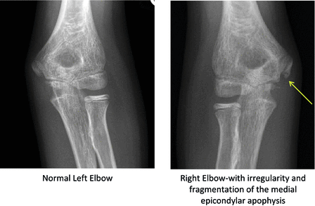
Fig. 7.1
Images from a 12-year-old pitcher with a 2-month history of medial elbow pain when throwing. (a) Normal left elbow. (b) Right elbow showing irregularity and fragmentation of the medial epicondylar apophysis
Treatment: The cornerstone of management of apophyseal injuries is relative rest. Pain should be used as a guide to the amount of activity that is acceptable. If the athlete is limping or has pain with walking, a walking boot, stiff-sole shoe, or crutches should be used until pain resolves and gait normalizes. Ice cup massage and over-the-counter pain relievers can be helpful for pain management. Physical therapy to work on improving flexibility and strength can be helpful. Modalities such as iontophoresis may be useful adjuncts to allow a safe return to sports. In milder cases a home exercise plan can be implemented instead of a formal physical therapy program. Surgical consultation is recommended for avulsion injuries with 2–3 cm of displacement.
Return to play: Young athletes may return to play when they are pain-free with activities of daily living and exercise, have full strength of the involved muscles, and have no evidence of an altered gait during or after activity. They should continue to perform their rehabilitation exercises to maintain strength and flexibility.
Osteochondrosis of the Elbow
Osteochondrosis is defined as “a focal disturbance of endochondral ossification” [58]. It has a multifactorial etiology, with no single factor accounting for all aspects of the disease. Etiologic factors include heredity, rapid growth, anatomic conformation, trauma, and dietary imbalances; however, only heredity and anatomic conformation are well supported by the literature [58].
Incidence: Osteochondrosis of the elbow, also known as Panner’s disease, occurs in young athletes between the ages of 5 and 10 years. It is more common in athletes who play sports that involve throwing (i.e., baseball) or weight-bearing on the upper extremities (i.e., gymnastics).
Mechanism of injury: It is believed to occur from an injury to the blood supply of the epiphysis in growing athletes. This leads to fragmentation of the capitellum and eventual resorption followed by reorganization of the epiphysis.
Signs and symptoms: Lateral elbow pain with activities, relieved by rest: Elbow stiffness and lack of full extension may develop, and possibly decreased pronation or supination. However, locking does not occur. On physical examination tenderness over the lateral elbow and capitellum is found [59].
Imaging: Radiographs will show flattening of the capitellum with irregular surfaces and radiolucent lesions. Over time (9–18 months) X-rays will show resolution of the condition with restoration of a rounded capitellar epiphysis.
Management: Treatment involves rest from aggravating activities and symptom control (i.e., pain management). Time alone will allow the epiphysis to revascularize and reorganize [59]. Repeat X-rays should be performed in 6–12 weeks to assess healing.
Return to play guidelines: The athlete may return to throwing activities or upper extremity weight-bearing activities when symptoms have resolved and there is radiographic healing of the capitellum. Plain radiographs may lag behind the clinical examination.
Prevention: Since the etiology is multifactorial, this condition may not be completely preventable. However, repetitive throwing and upper extremity weight-bearing are modifiable, so pitch count recommendations and general guidelines for periodic rest from sport should be followed.
Clinical pearls: It is important to differentiate this condition from osteochondritis dissecans (OCD) of the elbow. OCD occurs in a slightly older age group, may result in loose body formation causing mechanical symptoms of locking, and has a very different treatment and prognosis.
Bone Stress Injuries
Table 7.3 lists common bone stress injuries and their key exam findings. General principles for BSI are discussed below.
Table 7.3
Common bone stress injuries of the extremities
Location | Exam |
|---|---|
Metatarsals | Focal TTP metatarsal shaft |
Media tibial stress syndrome | TTP spans several cms of the posteromedial tibial border |
Tibia | Focal TTP, most commonly at the junction of middle and distal thirds of tibial shaft |
Fibula | TTP of fibular shaft |
Physis | Exam is usually unremarkable |
Proximal humerus | Pain occurs only with throwing |
Distal radius | Pain occurs with weight-bearing on the wrist |
Incidence: Stress reactions and stress fractures, known collectively as BSI, occur in young athletes. Prevalence is sport specific. A study of Australian track and field athletes reported that 20 % developed a stress fracture [60], while among US collegiate track and field athletes, 16 % developed a BSI [61]. A study at a national tennis training center demonstrated that 12.9 % of players had a stress fracture [62]. Importantly this study found that junior tennis players were more likely to have these injuries than adult players. In another study, BSI in adolescent and exercising women (mean age 18 y/o) were 30–50 % more likely when risk factors for female athlete triad were present [63].
Mechanism of injury: BSI may occur along a spectrum. Initially, periosteal and bone marrow edema occurs without cortical involvement. Stress reactions may progress to involve the cortex resulting in a stress fracture. Such injury may occur in otherwise healthy bone exposed to high-volume and biomechanical loads. Non-traumatic fractures occurring with modest biomechanical load or volume suggest that the bone is unhealthy (from diminished bone density or other metabolic factors). Complications of BSI include nonunion or complete fracture. Specific anatomic regions, such as the femoral neck, the anterior cortex of the tibia, and the metaphyseal/diaphyseal junction of the fifth metatarsal, have an increased propensity for nonunion in part based on total stress, and vascular supply.
Imaging: X-rays are recommended for any young athlete suspected of having a BSI. The sensitivity of plain radiographs may be as low as 10 % if done very early. A negative X-ray does not exclude BSI. X-rays may be repeated, though may never reveal the injury. If X-ray is non-confirmatory, additional imaging such as MRI should be considered. Sensitivity and specificity of BSI are highest for MRI and may allow for better staging of some BSI [61]. MRI staging includes grades 1–4, with grade 1 having mild periosteal edema only on T2 images with no fracture line progressing to grade 4 with severe periosteal edema on T2 images, marrow edema on T2 and T1 images, and a visible fracture line [61]. Other imaging modalities, including bone scan and CT scan, are falling out of favor for initial diagnosis as they expose young athletes to higher amounts of radiation.
Management: While management of BSI should be individualized, general principles of treatment include reduction of impact loading to allow for healing. Immobilization, protected weight-bearing (e.g., use of a walking boot, or long pneumatic splint), or non-weight-bearing with crutches may further reduce loading and control pain occurring with routine walking and weight-bearing. Cross-training with nonimpact activities such as biking or swimming to maintain cardiovascular fitness is recommended.
Return to play: When pain resolves, there is adequate bone healing, and no tenderness over the area of the stress fracture, the athlete may begin weight bearing activities followed by controlled impact and sport specific training. Return to play should occur with a structured on-field or on-court progression with intermittent participation and gradually increasing volumes. In a prospective study of collegiate track and field athletes, higher grade BSI were associated with a longer return to play [61]. Specifically, grade 1 and 2 injuries returned at approximately 13 weeks, while grade 3 and 4 injuries returned at approximately 24 weeks.
Prevention: Young athletes should have appropriate caloric intake, as well as calcium and vitamin D intake. Girls who develop menstrual dysfunction should be assessed for the female athlete triad, caloric insufficiency, and/or overtraining. All young athletes with recurrent BSI should be considered for evaluation for underlying bone metabolic disorders. Training volume should be carefully monitored. A gradual increase in training is necessary to prevent re-injury.
High-Risk Bone Stress Injuries
Overuse injuries may not be benign. Some present significant treatment challenges, with the potential to alter an athletic career and cause long-term health consequences. These injuries are listed in Table 7.4 and discussed in detail below.
Table 7.4
High-risk bone stress injuries of the extremities
Location | Exam | Imaging |
|---|---|---|
Femoral neck-tension side | Pain with passive hip internal rotation | Frequently only seen on MRI |
Anterior tibial cortex | Focal TTP of anterior tibia | X-ray may reveal the defect of anterior tibial cortex (“dreaded black line”) |
Tarsal navicular | TTP over mid-dorsal navicular (“N” spot) | MRI or CT |
Fifth metatarsal at metaphyseal/diaphyseal junction (“Jones fracture”) | TTP at proximal fifth metatarsal | May see cortical defect on X-ray. MRI if X-ray negative |
Femoral Neck Stress Fracture
Mechanism of injury: BSI occurs due to repetitive and rapid increases in training, or underlying bone health deficiencies.
Signs and symptoms: There should be a high index of suspicion for this injury in athletes who present with anterior hip pain or groin pain with running or other weight-bearing activities. Pain with a single-leg hop localizing to the groin is concerning. Pain with passive internal rotation or resisted external rotation may also be present.
Imaging: Hip X-rays should be performed with attention paid to the femoral neck for periosteal reaction or lucency. Since X-rays are frequently negative, MRI should be performed to determine if there is bone marrow edema, cortical involvement, or a fracture line. The location of a femoral neck stress fracture is critical to determining its appropriate treatment.
Management: Compression-sided (inferior) femoral neck stress fractures without displacement can be treated non-operatively with non-weight-bearing on crutches for approximately 6 weeks. Cross-training involving no weight-bearing may be considered prior to 6 weeks if there is no pain. Patients with tension-sided (superior) femoral neck stress fractures should be strictly non-weight-bearing and promptly referred to an orthopaedic surgeon for possible surgical fixation, since these stress fractures are at risk for nonunion and progression to complete fracture [65].
Return to play guidelines: For compression-sided injuries, after 6 weeks of non-weight-bearing, limited weight-bearing exercises may be introduced, followed by gradual progression to full weight-bearing exercises over another 6-week period. Repeat MRI is recommended by the authors to ensure resolution prior to full impact training.
Prevention: Similar to that described above for other BSI.
Clinical pearls: A high index of suspicion is critical to a timely diagnosis. Any athlete who develops anterior hip or groin pain in the setting of high training volumes should be removed from weight-bearing exercise and evaluated for a femoral neck stress fracture.
Stress Fracture of the Anterior Tibial Cortex
When treating exertional leg pain in young athletes, clinicians should be aware of a higher risk tibial stress fracture involving the anterior tibial cortex. These injuries occur on the tension side of the tibia and are notoriously difficult to treat. Tenderness on exam is localized to the anterior tibia, as opposed to the posteromedial tibial border in low-risk tibial BSI. Lateral radiographs may demonstrate a defect of the anterior tibial cortex (referred to as a “dreaded black line”) (Fig. 7.2). If X-ray is negative, MRI or CT scan with thin cuts should be obtained [66]. Limited weight-bearing and immobilization with pulsed electrical stimulation for 3–6 months have been described [67]. However, even with prolonged non-weight-bearing healing is not predictable. Therefore, surgical consultation is recommended. Surgical treatment with intramedullary nailing has been successful in adolescents, with an average of 4 months for return to sports [68].
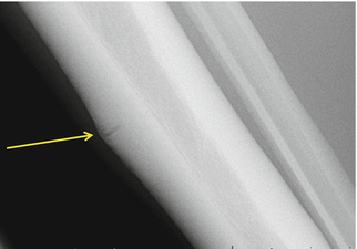
Fig. 7.2
“Dreaded black line”: Stress fracture of the anterior tibial cortex
Navicular Stress Fracture/Reaction
Incidence: Incidence of tarsal navicular stress fractures in young athletes has not been documented; however it was the most common lower extremity stress fracture in elite tennis players at a national tennis training center, with junior players being at greatest risk [62]. A case report of a 13-year-old female cross-country runner has also been reported [69].
Mechanism of injury: Increased load and stress to the medial mid-foot with excessive training. The midportion of the navicular has the higher risk, because this area is relatively avascular.
Related posts:
 Epidemiology of Injury in Elite Youth Sports
Injury Research in Pediatric and Adolescent Sports
Epidemiology of Pediatric and Adolescent Injury in Adventure and Extreme Sports
Epidemiology of Injury in High School Sports
Back Pain in the Young Athlete
Acute Lower Extremity Injuries in Pediatric and Adolescent Sports
Epidemiology of Injury in Elite Youth Sports
Injury Research in Pediatric and Adolescent Sports
Epidemiology of Pediatric and Adolescent Injury in Adventure and Extreme Sports
Epidemiology of Injury in High School Sports
Back Pain in the Young Athlete
Acute Lower Extremity Injuries in Pediatric and Adolescent Sports
Stay updated, free articles. Join our Telegram channel

Full access? Get Clinical Tree


