Fig. 2.1
The mechanism of induction for cancer-induced bone pain. The acidic microenvironment created by activated osteoclast-stimulated nocicepotors, which results in skeletal pain as the mechanism observed for cancer-induced bone pain
2.2 Assessment of Pain-Like Behavior in Ovariectomized (OVX) Mice as a Model of Postmenopausal Osteoporosis
Skeletal pain in OVX mice as a model of postmenopausal osteoporosis and that in OVX mice treated with BP were evaluated to elucidate mechanisms causing osteoporosis-related bone pain. OVX mice reveal osteoporotic changes 6 weeks after the surgery, of which total femoral bone mineral density (BMD) was significantly lower in the OVX group than in the sham group. Serum tartrate-resistant acid phosphatase 5b (TRAP5b), a bone resorption marker, was significantly higher in the OVX mice compared with the sham mice 4 weeks after surgery.
2.2.1 Evaluation of Pain-Like Behavior and Bone Metabolism in OVX Mice
2.2.1.1 Assessment of Pain-Like Behavior
Behavioral tests were conducted just after OVX and every 2 weeks thereafter. Thermal nociceptive testing (paw-flick test ) was conducted using an analgesimeter (Plantar test 7370, Ugo Basile, Italy). The mice were unrestrained, and radiant heat was applied to the plantar surface of the left hind paw until it was actively withdrawn by the animal. Paw withdrawal latency (PWL) was considered to be an index of the thermal nociceptive threshold, and a decrease in this measurement indicated thermal hyperalgesia [14]. Mechanical allodynia (von Frey test ) was measured using von Frey monofilaments on the left hind paw. The von Frey withdrawal threshold was determined by adjusting the stimulus intensity between 0.2 and 10 g equivalents of force and was estimated using the Dixon nonparametric test [15]. Motor functions were measured using the rotarod test, a test associated with pain-like behavior. The mice were placed on a cylinder, and the speed of rotation was set to 32 rotations/min. Latency to the fall from the rotarod was recorded. Lower values on these behavioral tests indicated greater skeletal pain [16].
2.2.1.2 Assessment of Skeletal Pain by Immunohistochemistry of Spinal Cord
We assessed skeletal pain by measuring c-Fos expression in the spinal cord, a functional marker of nociception [17]. The c-fos proto-oncogene is an immediate early gene and is expressed in response to both noxious and non-noxious stimuli [18, 19]. The L4–L5 segment of the spinal cord was immune-stained with c-Fos, and the number of c-Fos-labeled neurons in laminae I–II of that was counted.
In addition, a 0.02 mg/kg of body weight dose of alendronate (Merck & Co., Inc., NJ, USA), a BP, was administered to OVX mice subcutaneously once per day for 2 and 4 weeks after surgery (OVX + BP) [20, 22]. Behavioral tests, serum TRAP5b measurements, and immunohistochemical tests were performed in the sham and OVX mice.
2.2.2 Pain-Like Behavior After OVX and the Influence of BP
OVX mice had lower pain threshold on the paw-flick test and von Frey test, 4, 6, 8, and 10 weeks after surgery, compared with the sham mice (Fig. 2.2a, b), and the rotarod test showed a lower threshold 6 and 8 weeks after surgery (Fig. 2.2c). OVX mice were treated with BP (OVX + BP) for 2 weeks. The pain thresholds of the OVX mice improved significantly in all tests after administrating BP, and these effects remained 4 weeks after discontinuing BP treatment (Fig. 2.2a-c). The decreased pain threshold value due to OVX was improved significantly 2 weeks after administering BP (OVX + BP). These effects maintained until 10 weeks after discontinuing BP treatment [22].
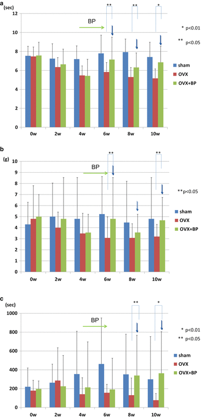
Fig. 2.2
The measurement of pain-like behavior. OVX mice showed a significant decrease in pain threshold value on the paw-flick test (a) and von Frey test (b) at 4, 6, 8, and 10 weeks after OVX and on the rotarod test (c) at 6 and 10 weeks, compared with the sham mice (*p < 0.05). The OVX-induced decrease was significantly improved at 2, 4, and 6 weeks after BP administration (OVX + BP). These effects were maintained until 4 weeks after the discontinuation of BP treatment. post-op (weeks), weeks after the surgery; BP (arrow), 2-week treatment with BP from 4 to 6 weeks after the ovariectomy (Figures were modified from Ref. [22])
The serum TRAP5b was significantly related to the pain threshold value, with correlation on the paw-flick test, on the von Frey test, and on the rotarod test. Furthermore, BP’s reduction in pain-like behavior was associated with decreased serum TPAP5b levels.
The number of c-Fos immunoreactive neurons, a marker of nociception, in laminae I–II of the dorsal horn of the spinal cord was significantly greater in the OVX mice than in the sham mice (Fig. 2.3). Those neurons were significantly lower in the OVX + BP mice than in the OVX mice (Fig. 2.3). These results, therefore, suggest that OVX mice with osteoporosis have greater skeletal pain than normal mice. In addition, the higher serum TRAP5b levels associated with OVX decreased significantly following BP treatment and were significantly correlated with pain threshold values [22], further suggesting that the skeletal pain accompanying osteoporosis is possibly associated with osteoclastic activity.
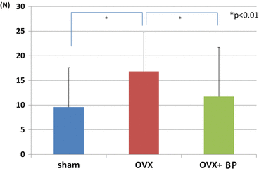
Fig. 2.3
Immunostaining for c-Fos in transverse sections of the L4–L5 level of the lumbar spinal cord. The number of c-Fos immunoreactive neurons in OVX mice (OVX) was significantly greater than that in both sham mice (sham) and OVX mice after BP administration (OVX + BP) (Figures were modified from Ref. [22])
Several studies show that estrogen decrease associated with OVX increases skeletal pain by augmenting nociceptive pathway excitability in the peripheral and central neuronal systems [23–25]. BP may also directly suppress the release of neurotransmitters or inflammatory mediators in the nervous system [26–29]. Estrogen decrease due to OVX may have caused skeletal pain by directly influencing the nervous system in our study. However, more than 80 % of intravenously administered BP is taken up by bone tissue within 24 h and remains in the tissue for a long duration [30]. Furthermore, our results suggest that the BP improvement of pain-like behavior was maintained even after discontinuing treatment for more than 4 weeks (Fig. 2.2). Therefore, the pain-like behavior associated with OVX and the inhibitory effects of BP are likely reflected by bone metabolic changes.
2.3 Mechanism of Skeletal Pain Induction Related to Osteoclast Activation
Osteoclasts degrade bone minerals by secreting protons through vacuolar H+-ATPases (V-ATPase), creating an acidic microenvironment [31, 32]. Acid causes pain through acid-sensing nociceptors [33, 34]. Two main classes of these nociceptors, TRPV1 and acid-sensing ion channels (ASICs) , are expressed in sensory neurons innervating the bone [34] and elicit pain signals when activated by acid stimuli [34, 35]. Previous studies of bone pain in a metastatic cancer model show that the acidic microenvironment created by bone-resorbing osteoclasts activates TRPV1 [9, 36, 37].
2.3.1 Acidic Environment in Bone by Activated Osteoclast and the Influence of BP
Histochemical findings in bone tissue demonstrated that the number of TRAP-positive osteoclasts in OVX mice was increased in comparison with that in sham mice (Fig. 2.4). In contrast, the number of TRAP-positive osteoclasts in OVX + BP was lower than that in OVX mice (Fig. 2.4). The V-ATPase expression was greater in OVX mice than that in sham or OVX + BP mice, respectively (Fig. 2.5). These findings, which osteoclast activity and V-ATPase expression were increased in OVX mice, indicated that the skeletal pain accompanying osteoporosis is possibly associated with the acidic microenvironment resulting from osteoclast activation [22].
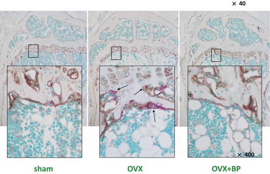
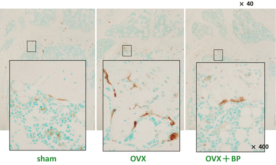
Fig. 2.4
Staining for TRAP in femora. Figures show double staining for localized ALP-positive osteoblasts (brown color) and TRAP-reactive osteoclasts (red) in the femoral trabeculae of sham, OVX, and OVX + BP mice. OVX mice demonstrate several TRAP-reactive osteoclasts (black arrow), while the other groups had a few flattened TRAP-positive osteoclasts (Figures were modified from Ref. [22])
Fig. 2.5
The histochemical detection of V-ATPase in femora. The immunoreactivity against V-ATPase (brown color). There are many V-ATPase-immunopositive osteoclasts on the bone surfaces in the OVX group, while only a few flattened immunoreactive osteoclasts can be seen in the sham and OVX + BP groups (Figures were modified from Ref. [22])
2.3.2 Involvement of Acid-Sensing Nociceptors in Skeletal Pain Induction Related to Osteoclast Activation
With regard to the induction of pain through acid-sensing nociceptors, two main classes of these nociceptors, transient receptor potential channel vanilloid subfamily member 1 (TRPV1) and acid-sensing ion channels (ASICs), are expressed in the sensory neurons innervating the bone [34, 35] and elicit pain signals when activated by acid stimuli [33, 34]. TRPV1 is a member of a family of polymodal and nonselective cation channels that are predominantly expressed by sensory nerve fibers and sensitized by protons [21] and improved the threshold [22]. ASICs are a major group of acid-sensing nociceptors and are expressed in sensory neurons innervating the bone and known to be related to the acid-induced pain [34, 35] in a similar manner as another important nociceptor TRPV1 [21, 22]. According to our study, the antagonists of TRPV1 or ASIC significantly improved the threshold value for pain-like behavior in OVX mice (Fig. 2.6) [22 and unpublished data]. In general, the secretion of protons by osteoclasts through V-ATPase is known as an acidification pathway during bone resorption [32]. We also demonstrated that a V-ATPase inhibitor significantly improved pain-like behavior in OVX mice (unpublished data). These results support our hypothesis that the acidic microenvironment produced by osteoclasts activates acid-sensing nociceptors and contributes to bone pain.
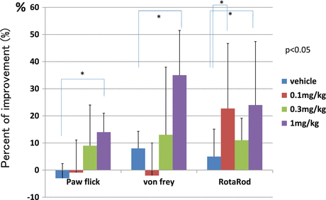
Fig. 2.6
Effects of a TRPV1 antagonist on pain-like behavior. All tests were conducted 6 weeks after OVX. A TRPV1 antagonist (SB366791), at a dose of 1 mg/kg, improved the threshold of pain-like behavior compared with the baseline value at 30 min after administration
2.3.3 Involvement of P2X Receptors in Skeletal Pain Induction Related to Osteoclast Activation
Extracellular adenosine triphosphate (ATP) and P2X receptors , which is a P2X receptor ligand, are implicated in nociceptive signaling under both normal and pathologic pain states. P2X receptors, which include seven subunits (P2X1–P2X7) belonging to the ligand-gated ion channel family, are activated by extracellular ATP [38, 39]. In the periphery, ATP can be released as a result of tissue injury, visceral distension, or sympathetic activation and can excite nociceptive primary afferents by acting as homomeric P2X3 or heteromeric P2X2/3 receptors [38]. Recent studies have demonstrated that the level of ATP is increased by osteoclast activation under a high bone turnover state in osteoporosis and that several P2X receptor subunits exist in bone tissue [40–42]. Particularly, many studies have demonstrated that P2X7 receptor expressed in osteoclasts or osteoblasts and had significant roles in bone homeostasis or in association with pathogenesis of osteoporosis [43–46]. However, there are relatively few studies concerning expression and function of P2X2 [45] or P2X3 receptors in the bone. A recent study has indicated that a P2X3 receptor could contribute to bone cancer pain through the upregulation of increased local ATP levels in bone tissue [42]. P2X3 receptor, a specific ATP-sensitive ligand-gated ion channel, is selectively localized on peripheral and central processes of sensory afferent neurons and participates in the role of nociceptive signaling [47, 48]. The P2X3 receptor is natively expressed as a functional homomer and as a heteromultimeric combination with the P2X2 receptor [47–49]. Based on the results of those studies, we also hypothesize that increased local ATP underlying a high bone turnover state in osteoporosis activates the P2X receptor in bone tissue, and those activated P2X2/3 might has a role in the induction of skeletal pain.
Related posts:
 Vertebral Strength Changes as Assessed by Finite Element Analysis
 Spondylosis and Osteoporotic Vertebral Fractures
 Applications of Teriparatide for Fracture Repair and Osteosynthetic Surgery in Osteoporosis
 Collagen Cross-Links as a Determinant of Bone Quality
 Sarcopenia and Osteoporosis
 Back Extensor Strengthening Exercise and Osteoporosis
Stay updated, free articles. Join our Telegram channel

Full access? Get Clinical Tree


