Osteochondritis Dissecans of the Knee


Osteochondritis Dissecans of the Knee


Frederick M. Azar

S. Terry Canale


Osteochondritis dissecans (OCD) of the knee is a common entity in both adults and children. Its incidence has historically been estimated to be between 0.02% and 0.03% based on knee radiographs, and 1.2% based on knee arthroscopy (5,11,27). More recent investigators have reported much higher frequencies (1,21). Osteochondritis dissecans occurs twice as often in males as in females. Osteochondritis dissecans is most common in the medial femoral condyle, followed by the lateral femoral condyle; its occurrence in the patella is rare. It primarily affects young athletic individuals and can significantly impair knee function and activity and lead to long-term disability (10).

The etiology of OCD is controversial and remains unclear. Theories include ischemia, repetitive microtrauma, familial predisposition, endocrine imbalance, epiphyseal abnormalities, accessory centers of ossification, growth disorders, osteochondral fracture, repetitive microtrauma with subsequent interruption of interosseous blood supply to the subchondral area of the epiphysis, anatomic variations in the knee, and congenitally abnormal subchondral bone.

It is important to distinguish OCD from acute osteochondral fracture (Fig. 34-1). In addition, an OCD lesion that has separated from a vascular bony bed should be distinguished from one that has separated from an avascular bony bed (osteonecrosis). Osteochondritis dissecans is more common in adolescents and young adults than is osteonecrosis.

Several classification schemes for OCD have been proposed. Bradley and Dandy (5) developed a classification for the knee and limited the term OCD to expanding concentric lesions of the medial femoral condyle that appear during the second decade of life and progress to concave, steep-sided defects. Cahill and Berg (6) proposed a classification scheme based on the appearance of the lesion on scintigraphy. Dipaola et al (12) classified lesions according to their appearance on magnetic resonance imaging (MRI) and associated specific findings with the potential for fragment detachment. Guhl (19) developed an intraoperative classification based on cartilage integrity and fragment stability noted at the time of arthroscopy.

Although the natural history of these lesions has not been conclusively defined, current evidence suggests that they persist and can lead to further cartilage degradation (32). Primary prognostic factors, in addition to the age of the patient, include progression, size, stability, amount of subchondral bone present, and location of the lesion, especially as it relates to weight bearing. Lateral femoral condylar lesions tend to be more posterior, larger, less stable, and more fragmented than medial lesions.

Although nonoperative treatment has a limited role in patients with symptomatic osteochondral lesions, multiple forms of nonoperative treatment have been described, including periods of immobilization, activity modification, and non-weight bearing. Prolonged immobilization should be avoided because joint motion affects articular cartilage attrition. Cahill et al (7) noted a 50% failure rate with conservative treatment in a juvenile population with OCD.

A number of operative procedures have been devised for treatment of OCD, ranging from simple debridement to autograft/allograft or chondrocyte implantation (Table 34-1). Arthroscopy is preferable to arthrotomy; however, it may be necessary to convert an arthroscopic procedure to an arthroscopic-assisted mini arthrotomy to obtain adequate fixation of an unstable lesion.


FIGURE 34-1

Unstable lesion that requires fixation.


INDICATIONS/CONTRAINDICATIONS

See Table 34-1 for surgical treatment options and their indications.

There are few contraindications to surgical treatment of OCD. Contraindications to specific surgical techniques may include the size of the lesion, with some procedures suited for only small (<2 cm2) lesions; the activity demands of the patient (high vs. low demand), and the age of the patient (osteochondral allo- and autografts, autologous chondrocyte implantation usually not indicated in patients with open physes). The location of the lesion also may be a contraindication to a particular technique. Patient willingness to comply with postoperative weight-bearing restrictions also must be considered.


PREOPERATIVE PLANNING


History and Physical Examination

The most common complaint of patients with OCD is pain, which usually is difficult to localize and typically is present for several months. Patients also may report swelling or mechanical symptoms such as catching and popping if the lesion is partially or completely separated. Approximately 80% of juvenile patients have symptoms for an average of 14 months before initial presentation. A history of trauma to the knee is given by 40% to 60% of patients (8). Examination also may reveal effusion, joint line tenderness or tenderness over the lesion, limitation of motion, a positive McMurray’s sign, and quadriceps atrophy. The patient may walk with an externally rotated gait to avoid contact of the medial femoral condyle with the medial tibial spine. A thorough history and physical examination are necessary to rule out other causes of knee pathology.


Imaging

Imaging techniques are important not only for diagnosis but for treatment decision-making. The size, location, and stability of the lesion are critical to choosing the appropriate technique.


Radiographs

Most OCD lesions can be easily identified on plain radiographs. Anteroposterior, lateral, notch or tunnel, and patellofemoral views are recommended. Most lesions occur in the lateral portion of the medial femoral condyle, and they are more clearly seen on the notch and lateral views. Lesions of the lateral femoral condyle tend to be more posterior than those in the medial
condyle (Fig. 34-2). The size of the lesion usually can be accurately determined on plain radiographs. Most small lesions are stable; lesions with a sclerotic margin of 3 mm or more and lesions larger than 0.8 cm2 have an increased risk of being loose. Comparison radiographs should be obtained in juvenile and adolescent patients because an OCD lesion may be confused with ossification centers, which may cause transient symptoms but usually resolve spontaneously within 6 to 12 months. In addition, OCD is bilateral in approximately 30% of patients (Fig. 34-3).


TABLE 34-1. Surgical Treatment Options for Osteochondritis Dissecans of the Femoral Condyle


Procedure


Technique


Indications


Comments


Arthroscopic debri dement


Shaving of loose chondral flaps


Small defects (<2 cm2) in areas of limited weight bearing


May provide short-term relief.


Internal fixation with K-wires, screws, biodegradable rods, or pins (arthroscopy or mini arthrotomy)


Removal of fibrous tissue from crater, antegrade or retrograde internal fixation. Cancellous graft from proximal tibia or femoral condyle can be added.


Unstable lesion, hinged or loose in crater.


Risk of hardware breakage or loosening, protruding hardware causing articular wear; second procedure may be needed for hardware removal.


Perichondral/periosteal graft


Graft harvested from distant (perichondrium) or adjacent (periosteum) site, implanted over defect to serve as joint buffer, source of mesenchymal stem cells


Small defect in non-weight-bearing area in young patient.


Long-term (5 to 10 year) results have been disappointing.


Abrasion chondroplasty/ microfracture


Arthroscopic drilling, microfracture with sharp awl, abrasion with a burr. Combined with removal of loose bodies and debridement of loose cartilage.


Creation of bleeding surface to access pluripotential stem cells from marrow.


Younger patient Normal alignment Mechanical symptoms (catching, locking, swelling) Lesion <2 cm2 with stable borders


Good initial treatment


Results in mainly fibrocartilage matrix formation, which has more type I collagen, cannot withstand compressive and shear loads as well; may degenerate over time.


Osteochondral autograft (OATS, COR, mosaicplasty)


Transplantation of single or multiple plugs from areas with less weight-bearing to fill defects in areas with more weight-bearing function.


Defect ≤2 cm2


Inability to treat large defects due to limited availability of donor cartilage.


Potential problems from donor site.


Osteochondral allograft


Transplantation of single or multiple plugs from fresh allograft femoral condyles


Defect >2 to 3.5 cm2 Isolated lesion Limited donor site Young patient


No morbidity from donor site, improved ability to shape and tailor graft. Risk of disease transmission.


Autologous chondrocyte implantation (ACI)


Cells obtained from arthroscopic cartilage biopsy, released from matrix and culture expanded, then placed in defect under periosteal patch.


Younger (15 to 55 years) patient Active patient Single or multiple defects 2 to 10 cm2


Ligamentous or meniscal lesions, joint malalignment, patellofemoral instability must be corrected; complete meniscal deficit may be contraindication.


COR, chondral osseous replacement; OATS, osteochondral autograft transfer systems.


Magnetic Resonance Imaging

Magnetic resonance imaging is recommended to determine the extent of the lesion. Common MRI characteristics include a high-signal-intensity line or cystic area beneath the lesion, a high-signal-intensity line through the articular cartilage, and a focal articular cartilage defect on T2-weighted spin echo and short tau inversion recovery (STIR) images
(Fig. 34-4). Currently, a spoiled gradient (SPGR) echo sequence using fat suppression and three-dimensional acquisition is considered the optimal technique for evaluating articular cartilage lesions. The presence of fluid around the fragment or focal cystic areas beneath the fragment is the best indicator of instability; the absence of a zone of high-signal intensity at the interface of the fragment and the bone is a reliable sign of lesion stability (28). Magnetic resonance imaging also is useful for identification of associated ligamentous and meniscal injuries.


FIGURE 34-2

Recommended radiographic views for evaluation of OCD of the knee. A: Anteroposterior. B: Lateral. C: Tunnel.


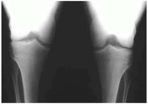
FIGURE 34-3

Bilateral OCD lesions of lateral femoral condyles.


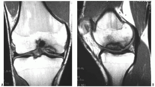
FIGURE 34-4

MRI evaluation of OCD. A: AP-coronal plane view. B: Lateral sagittal plane view.


Computed Tomography Scanning

Computed tomography (CT) scanning can delineate the bony contours and allow determination of the amount of cortical bone present in the osteochondral fragment (8). Healing potential is determined by the quantity of bone attached to the major fragment, and healing can be followed with tomograms until union.


Bone Scanning

Bone scanning (technetium bone scintigraphy) may be helpful to determine the extent of activity within the lesion and to monitor progress of healing (6,37). This has been shown to be more reliable in patients with open physes than in skeletally mature patients.


SURGERY


Patient Positioning

The patient is placed supine with the prepared and draped limb angled off the lateral aspect of the table; a leg holder or lateral post can be used.


Technique

Jun 13, 2016 | Posted by in ORTHOPEDIC | Comments Off on Osteochondritis Dissecans of the Knee

Full access? Get Clinical Tree

Get Clinical Tree app for offline access