Fig. 9.1
X-ray showing an os trigonum
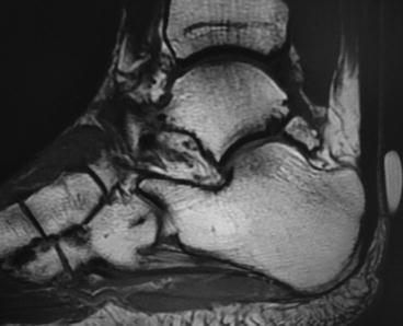
Fig. 9.2
MRI of os trigonum
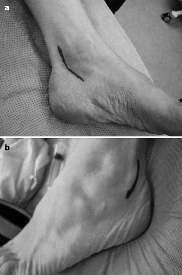
In recalcitrant cases of symptomatic os trigonum injuries, surgical excision can result in good relief. Open surgical approach is typically posterolateral, unless preoperative imaging reveals that the ossicle or fracture is displaced medially.2 Arthroscopic excision is described elsewhere in this text but has recently been proven to have good results in athletes.3 The patients can be placed prone to get better visualization with both open and arthroscopic excision, and in case another incision can be made, otherwise, lateral position is preferred. Some authors describe surgical extirpation of the bone arthroscopically as well, which is explained elsewhere in this text. The typical lateral open approach gains access to the posterior ankle and subtalar joint, when making the incision just behind the peronei (Fig. 9.3). This avoids the posterolateral neurovascular structures. The peronei are retracted anteriorly and a posterior capsulotomy is performed. A rongeur is often needed to remove the associated posterior synovitis. Care is taken to protect the FHL medially. A small osteotome can be used to free up the posterior ossicle or fracture talar process. Dorsiflexion and plantarflexion of the foot can help identify the posterior joints and the offending ossicle. It should be removed in toto (Fig. 9.4). The posterior talus is smoothed with a rasp, particularly the portion which comes in contact with the FHL. The tendon itself is inspected for any tears or stenosing tenosynovitis, which can be repaired or debrided as needed. Potential complications are excessive scarring and fibrosis of the FHL tendon and neurovascular injury. The area of resected bone can be covered with bone wax. The posterior articular surfaces are inspected for chondral defects and smaller loose bodies. Also, 1 cc of dexamethasone phosphate can be injected to decrease any adhesions. The capsule and peroneal retinaculum is re-approximated with absorbable suture. If additional lateral ankle procedures are needed, the incision can be extended anteriorly. Skin is closed with nonabsorbable suture. Patients can weight-bear post-surgery with a cast boot, generally for 4 weeks unless other procedures dictate otherwise. Formal physical therapy is initiated at 4 weeks. Patients can return to sports and dance between 6 and 12 weeks.