Orthopedic trauma
P.Christopher Metzger and Mark Lombardi
Introduction
As the average age expectancy approaches 80 years in the United States of America, musculoskeletal injuries can be expected to increase in number. This statement takes on significant ramifications when coupled with the knowledge that an estimated 78 million ‘baby boomers’ are either in their seventh decade or rapidly approaching it. The baby boomer population consists of anyone born between 1946 and 1964 according to the US Census Bureau. Today, more than ever, many geriatric patients lead very active and productive lives. Unfortunately, such a lifestyle can be dramatically affected by an inadvertent slip or fall that may produce an orthopedic injury. As these injuries become more prevalent they can be expected to have a profound effect on both society and its already stressed healthcare system. This chapter focuses on the rehabilitation of such orthopedic injuries in the geriatric population.
Rehabilitation may be defined as the restoration of normal form and function after an injury or an illness (Dirckx, 2001). What is meant by ‘normal form and function’ varies from individual to individual. The desired goal for the injured patient is to return them to their preinjury activities, knowing that this may not always be possible.
Fractures of the proximal femur are a common and increasing cause of hospitalization. In 2004 there were more than 320 000 hospital admissions necessitated by hip fractures in the US. It is projected that there will be more than 500 000 hip fractures by 2040 (CDC, 2010). At a cost of approximately $27 000 per patient one can see the staggering economic burden produced by such injuries. Roughly 4% of all deaths from injury in the US are caused by hip fractures (Bergen et al., 2008). In the geriatric population hip fractures are usually associated with low energy trauma. Death may occur when there is exacerbation of medical comorbidities (e.g. diabetes mellitus, coronary artery disease, chronic obstructive pulmonary disease etc.) caused by the resulting immobility or postoperative complications. Only 25% of these patients will make a full recovery, 30% will require nursing home care and 50% will require the use of a cane or walker. Within approximately 12 months, 30% of these patients will die (Moran & Wenn, 2005). Data from Europe indicate that hip fractures pose the same problems (Lippuner et al., 2005). Such statistics point out the need for expert and efficient musculoskeletal care.
The goal of fracture care in this age group is early mobilization and the eventual restoration of function. Prolonged periods of immobility increase the risk of deep vein thrombosis (DVT), pulmonary embolism, pressure ulcers, pneumonia and joint contractures. Surgical intervention, when necessary, is best performed within 48 hours after injury if the patient has been deemed medically stable. The patient, family, orthopedic surgeon and all those who provide ancillary services must understand that often there is a decrease in the healing potential of a geriatric patient. (See Chapter 60, Fracture Considerations.)
Basic principles for rehabilitation
The goals of rehabilitation are to: (1) control and reduce inflammation, (2) restore motion, (3) develop motor control and coordination, (4) regain strength and (5) restore function. The rehabilitation process in pelvic or lower extremity injuries is begun by mobilizing the patient. This consists of getting the individual out of bed into a chair and is followed by ambulation with external support (cane, crutches or walker). At the same time, joint mobility and flexibility must be restored. This is accomplished with active, active-assisted and passive range of motion (ROM) exercises. As both mobility and ROM are regained, emphasis must be placed on reacquiring strength, joint stability, motor control, proprioception and coordination.
It is desirable to start mobilization as soon as the patient’s medical condition permits. Learning to ambulate with either crutches or a walker in a partial weight-bearing fashion is a challenge for most. The amount of energy required to perform limited weight-bearing is 30–50% greater than that required for normal ambulation (Hsu et al., 2008). This added demand can be particularly taxing for the elderly individual, especially if there is a decreased cardiopulmonary reserve.
In upper extremity injuries, patient mobilization is usually not as difficult to attain. The only necessary instructions may be to keep the arm elevated and to educate the patient on how to get in and out of bed and chairs without putting pressure on the injured extremity. In general, the more severe injuries, those with upper and lower extremity involvement, pose a greater obstacle to mobilization. In such instances initial attention may have to focus on simple transfers from bed to chair because ambulation may not be possible. The use of adaptive equipment, such as forearm supports on assistive devices, may prove to be necessary. The geriatric patient may already have some pre-existing impairment of mobility that has to be taken into consideration. The goal of rehabilitation is to get the patient back to their preinjury status if at all possible.
Motion
One of the therapist’s responsibilities is to instruct and assist the patient in the restoration of ROM after injury. At all times, the physician should communicate with the therapist regarding any precautions or restrictions. Such communication should take place on a regular basis and must always be documented.
Motor control and coordination
Motor control is necessary before any active exercises can begin or progress. Sometimes electrical stimulation is needed to activate muscles that demonstrate atrophy, muscle inhibition or painful muscle guarding (Hopkins & Ingersoll, 2000). Coordination is crucial to motor control. It involves smooth and accurate movement of the joints in the kinetic chain. The timing and sequencing of movements of ipsilateral and contralateral joints requires neural control and musculoskeletal integrity. For example, a humeral fracture that disrupts the coordinated movement of the involved arm also reduces the contralateral arm swing during normal reciprocal gait. Proper breathing, decreased muscular guarding and reduced abnormal flexor and adductor tone in either the upper or lower extremities facilitates improved muscle activity and coordination.
Strengthening
When some degree of comfortable motion and muscle control are attained, strengthening can be started. Increased strength often results in increased motion. It has been shown that age is no barrier to regaining or even increasing strength. (See Chapter 61, Contractures and Stiffness.)
An effective method of strengthening is progressive resistive exercise (PRE). Initial efforts with strengthening may begin with isometric training. Isometric strength training may provide increased joint stability, promoting improved joint motion, as ROM increases. As the patient’s strength increases, the therapist may elect to progress strengthening through techniques that exercise each muscle group with enough resistance to allow 20–30 repetitions. It is recommended that the therapist break the patient’s sets/reps into more manageable bouts of exercise to prevent fatigue that often can lead to poor performance or injury. Once 30 repetitions can be achieved, the resistance is increased and the progression of repetitions from 20 to 30 is repeated. Another method involves the patient completing three sets of 10–15 repetitions, decreasing the resistance with each set, or three sets using the same resistance but decreasing the number of repetitions (from 20 to 15 to 10).
Adaptation
At some point during rehabilitation it may become evident that there will be some permanent functional limitation or disability. Changes in anatomy and consequently in function, resulting from the injury, may force changes in the patient’s movement patterns thus affecting their lifestyle. In order to adapt to these changes different training techniques or equipment may be needed. These needs may be apparent early in the rehabilitation period if, for instance, there has been a major amputation. In other cases, it may become evident later in the course of rehabilitation that permanent loss of joint motion or strength is inevitable and that compensation during work or play is required. Loss of joint motion during the rehabilitation period should be communicated with the physician on an ongoing basis in an attempt to minimize the degree or severity of loss.
The ability to restore some form of useful activity in the involved extremity is one of the primary goals of rehabilitation, although it may not always be attainable. It may be that the patient will need to adjust to a more sedentary lifestyle or pursue activities that are less physically demanding. Learning to accept these limitations is part of regaining a meaningful life.
Treatment of osteoporosis
Osteoporosis is a metabolic bone disease characterized by decreased bone mass and bone quality. Changes in bone mineral density (BMD) and quality increases the patient’s risk of fragility fractures. Osteoporosis is a ‘silent’ condition that is generally asymptomatic until a fracture occurs (Sinaki, 2003). It is defined by a BMD that is 2.5 or more standard deviations below that of normal young adults (WHO, 2003). In the US it has been estimated that 4–6 million women and 1–2 million men greater than 50 years of age have osteoporosis. In 2005 the cost for treating osteoporotic fractures in the US was $17 billion and is expected to increase by 50% by 2025 (Lim et al., 2009). It can therefore be seen that early detection and treatment of osteoporosis would play an essential role in decreasing healthcare costs. (See Chapter 18, Osteoporosis and Spine Fractures.)
There have been promising results in the treatment of osteoporosis and osteopenia through calcium supplementation, adequate vitamin D intake and the use of biphosphonates, estrogen and progesterone (Hormone Replacement Therapy – HRT) (Martin, 2012), calcitonin, teriparatide and testosterone. Pharmacological intervention is not without risk and the therapist should work with the patient, physician and nutritionist to insure that the patient is monitored and progressed appropriately (Martin, 2012). If possible, regular physical activity, including weight-bearing and resistive exercise, may also be helpful if introduced and advanced appropriately following recommended guidelines to safe exercise in patients with osteoporosis/osteopenia (Martin, 2012).
Rehabilitation after specific injuries
Fractures of the proximal humerus
Fractures of the proximal humerus are the third most commonly encountered fractures in the geriatric population, with only hip and distal radius fractures seen occurring more often. The frequent occurrence of this fracture in the elderly population certainly suggests an association with osteoporosis and impaired balance. A fall onto an outstretched hand (FOOSH) is the most common mechanism of injury for these fractures. Fortunately approximately 85% of these fractures are either nondisplaced or minimally displaced (Flynn, 2011). These fractures can be treated by sling and swathe immobilization for 10–14 days followed by gentle ROM exercises. Elbow flexion and extension as well as forearm pronation and supination can be started during this period of immobilization. Prior to initiating the exercise program for the shoulder, clinical continuity (the fracture moves as a single unit) must be present.
If there is stability at the fracture site, passive and active assisted ROM is started early. This consists of pendulum exercises and supine external glenohumeral rotation with a stick. About 3–4 weeks after the fracture has occurred, active-assisted forward elevation, pulley exercise, extension and isometrics can be added (Withrow et al., 2010). After this first phase has been complete, active and early resistive exercises become important. Therabands are often used to strengthen the shoulder rotators and deltoid muscle. A program that emphasizes further stretching and strengthening is appropriate 3 months after fracture.
It is important in the early stages of fracture healing for the patient to avoid using the affected arm when getting into and out of bed or a chair. Such actions can displace the fracture even when there has been stable internal fixation. Displacement is more likely to occur in the patient with multiple injuries or limited cognitive capabilities. Throughout the entire rehabilitation program, the therapist should be working with and assessing functional mobility of the cervical spine, scapula, elbow, wrist and hand. Most patients with fractures of the proximal humerus do obtain satisfactory results; however, it must be understood that usually there is some resultant loss of motion and strength. Complications include malunion, delayed union, nonunion, loss of motion, stiffness and post-traumatic arthritis.
Open treatment may prove to be necessary when a displaced fracture cannot be reduced. Treatment options consist of either a closed reduction with percutaneous pinning, an open reduction with internal fixation using a precontoured locking plate or hemi-arthroplasty when the articular surface is non-reconstructable.
The results of open reduction with internal fixation for proximal humerus fractures demonstrate good surgical outcomes when correct surgical technique is employed. Brunner et al. (2009) reported that the most common postoperative complication was screw perforation into the glenohumeral joint. Primary hemiarthroplasty is usually successful in eliminating pain; however, in many instances only moderate function and poor strength levels were achieved. It is felt that the reduction in function is due to lack of rotator cuff integrity (Gronhagen et al., 2007).
Fractures of the distal radius
Distal radial fractures account for approximately 20% of all fractures and therefore are the most common fractures seen in the upper extremity. Such fractures usually occur following a FOOSH and are seen quite often in the geriatric population, particularly women with osteoporosis (Flynn, 2011). A Colles’ fracture involves the distal radial metaphysis and demonstrates dorsal angulation and displacement. Often there may be comminution and intraarticular extension of the fracture. A Smith’s fracture demonstrates volar angulation of the distal radius with resultant instability.
Treatment options for these fractures include simple cast immobilization, closed reduction with cast application, closed reduction with external fixation and open reduction with internal fixation. The trend today is toward open reduction and internal fixation using a volar fixed-angled locking plate for displaced intraarticular fractures. The usual indications are radial shortening of more than 3 mm, dorsal tilt greater than 10° or intraarticular displacement of more than 2 mm (American Academy of Orthopedic Surgeons, 2011). The possible complications that may result from this particular type of fracture include delayed union, nonunion, malunion, median nerve compression, tendon damage, post-traumatic arthritis and loss of motion.
Restoration of motion and strength, which leads to improved function, is of vital importance in the rehabilitation of these fractures. Early rehabilitation includes management of pain and edema, digital ROM and care of the surgical site (Smith et al., 2004). After appropriate consultation with the attending physician, ROM and strengthening exercises for areas of the upper extremities that are not immobilized should be initiated immediately, if possible, to prevent residual stiffness in the shoulder, elbow and hand. Once immobilization is no longer necessary, active-assisted and active exercises are encouraged in all six directions – flexion, extension, radial and ulnar deviation, pronation and supination. Modalities such as hydrotherapy, electrical stimulation, heat, cold, or ultrasound may be helpful. Depending on the status of fracture healing and the amount of stiffness, the therapist may incorporate specific mobilization techniques to increase the ROM. The initiation of muscle control and coordination may be difficult when motion is painful. In addition to addressing the inflammatory process, which causes pain and swelling, the therapist should attend to head, neck and trunk posture, which may contribute to the patient’s discomfort and limitation of movement. As ROM in the wrist increases, strengthening exercises using motion against resistance should be included in the treatment plan. The final goal should be to restore ROM and strength of the injured wrist to preinjury levels. Unfortunately this is not always possible and some degree of impairment may remain as a result of the fact that normal architecture could be restored secondary to the severity of the injury (Smith et al., 2004).
When rehabilitating a patient who has undergone an open reduction with internal fixation using a volar plate, attention must be directed accordingly to prevent a contracture of the pronator quadratus, which could cause a limitation of forearm rotation.
Intertrochanteric fractures
An intertrochanteric fracture occurs along the line that is located between the greater and lesser trochanters. These fractures are seen most commonly in the elderly and are usually the result of a fall. The goal of care for this particular type of fracture should be to restore the patient to his or her preinjury status as quickly as possible. The potential benefits of operative intervention include rapid mobilization, ease of nursing care, shorter hospitalization, decreased mortality and restoration of function.
Stable intertrochanteric fractures are still best treated with a sliding screw (Flynn, 2011). The treatment of the unstable intertrochanteric fracture remains somewhat controversial but often requires the use of a cephalomedullary nail. The surgery should be performed within the first 48 hours if at all possible. The success of the surgical procedure largely depends upon: (1) bone quality, (2) fracture pattern, (3) accuracy of the reduction and (4) the adequacy of internal fixation.
The major goal of rehabilitation after an intertrochanteric fracture is to enable the patient to walk, especially if they were ambulatory prior to the injury. Mobilization of the patient should be initiated almost immediately after completion of the surgical procedure. ROM exercises are encouraged as soon as the initial pain subsides and the patient can safely cooperate with the physical therapist. ROM in all directions is advised, to prevent flexion and adduction contractures that can make ambulation more difficult. Getting the patient to a level where they can control the involved limb is essential to permit adequate mobility in bed, preventing and/or lessening the occurrence of pressure ulcers, to allow for independent transfers into and out of bed and to promote the initiation of weight-bearing and restoration of gait (Kagaya & Shimada, 2007). Balance and coordination instructions are given concurrently with all phases of rehabilitation.
Usually the patient should get out of bed and transfer to a chair on the day following surgery. With intertrochanteric fractures, partial weight-bearing may often be necessary. The weight-bearing status is determined by the accuracy and stability of the reduction achieved at the time of surgery, bone quality, premorbid status and mental alertness. The patient who is not strong enough to manage partial weight-bearing or not coherent enough to understand the therapist’s instructions may be limited to a wheelchair and/or pre-gait activities, such as sit to stand and static stance with weight shift until they are stronger or until the fracture has healed sufficiently to permit full weight-bearing. Early assisted swing (slide) phase of gait of the involved leg may be helpful in facilitating proper weight-bearing and restoration of functional gait in the future. This is achieved with the patient standing in parallel bars or with a walker, and simply sliding the foot of the involved leg forward and backward or lifting the involved leg over a lower obstacle (cane, cup or cone) that is placed on the floor in front of the patient.
Strengthening the hip abductors gradually reduces the Trendelenburg gait pattern commonly seen following a hip fracture. PREs are used, starting with abduction while standing or the use of a sliding board while supine. As strength increases, the patient is instructed to perform the exercises while lying on the contralateral side, thus abducting against gravity. Coexisting musculoskeletal and cardiovascular conditions may necessitate modification of these positions. Once 20–30 repetitions can be performed, progressive resistance is added. Strengthening the hip flexors, extensors, rotators and adductors is also important to insure restoration of muscle strength, flexibility and endurance to allow progression of gait.
After the fracture heals and rehabilitation is complete, occasional decreased mobility may be the end result. Patient adaptation may involve having to accept the permanent use of a cane or walker to aid in balance, reduce Trendelenburg characteristics associated with weak abductors seen in gait, and to increase patient confidence, safety and mobility. The patient with an intertrochanteric fracture should be expected to transfer and ambulate independently before being discharged home. If this is not possible, placement in an assisted living facility or a nursing home may be necessary.
Femoral neck fractures
Femoral neck fractures occur most commonly in the eighth decade of life as a result of bone that is weakened by either osteoporosis or osteomalacia. The most common mechanisms of injury are either a fall that causes a direct blow to the greater trochanter or forced lateral rotation of the lower extremity (Bucholz et al., 2009). If the fracture is displaced, often the arterial supply to the proximal end of the femur is disrupted, thus creating an environment favorable to the development of either a nonunion or avascular necrosis.
Elderly patients with nondisplaced or valgus impacted fractures can be treated with percutaneous pin fixation using cannulated screws (Flynn, 2011). The displaced fracture is best treated with hemiarthroplasty using a cemented stem. Today a trend is developing that suggests perhaps total hip arthroplasty is favorable over simple hemiarthroplasty for the highly functional elderly patient (Flynn, 2011).
If a posterior approach is used when either a hemiarthroplasty or total hip arthroplasty is performed, caution must be used for the first several weeks in order to prevent hip dislocation (see Chapter 21, Total Hip Arthroplasty). A dislocation may occur with a combination of excessive hip flexion, adduction and internal rotation. The avoidance of this position in the early postoperative period is imperative. Safety measures must also be taken when patients are putting on their stockings and shoes, recumbent in bed, sitting upright or rising from a chair or recliner. In cases in which the stability of the prosthesis is in question, an abduction pillow is extremely helpful. The patient should also be instructed to sit in a lean-back chair so that the hip is flexed no more than 90°. In most cases, when a prosthesis is inserted, a graduated weight-bearing program is indicated.
Recent studies suggest that the therapist work in concert with the medical team to address changes in affect identified in patients having sustained a femoral neck fracture (Olofsson et al., 2005).
Supracondylar fractures of the femur
The supracondylar region of the distal femur is often weakened by osteoporosis and thus even low energy forces can create complex fracture patterns. The resulting fractures are often comminuted, displaced and intraarticular, making management quite difficult. Years ago treatment consisted of traction and cast bracing, which unfortunately often resulted in a loss of joint motion. Today internal fixation techniques (intramedullary nailing or plate fixation) have been developed that allow for an anatomical reconstruction of the distal femur, more rigid internal fixation and earlier patient mobilization, thus allowing for improved ROM and function. Indications for nonoperative treatment include nondisplaced fractures, fractures occurring in patients who are not candidates for surgery, and fractures in patients who do not ambulate.
If rigid internal fixation has been achieved at the time of surgery, the use of a continuous passive motion machine may often prove to be quite helpful. This encourages increased motion, less postoperative swelling and reduces the incidence of quadriceps adhesions. For the first 6 weeks partial weight-bearing with a walker is allowed only if stable fixation is present. At the 6-week point, weight-bearing can be increased to tolerance providing there is radiographic evidence of healing. Full weight-bearing with external support is often not possible until the 12th week. In instances in which stable fixation has not been achieved supplemental support with a cast brace may prove to be necessary.
The same principles that govern motor control, coordination, strengthening and adaptation in fractures of the proximal femur apply to fractures of the distal femur. In addition, attention to distal lower extremity pain, weakness and decreased ROM will be required. The therapist should be aware of and proactive in the management of any patient having weight-bearing restrictions and decreased mobility issues as a result of lower extremity fracture and surgery with regard to an increased incidence of DVT or pulmonary emboli (PE). While Homan’s Sign is but one tool that the therapist has at their disposal to assess for DVT, they need to be aware that the test has a low sensitivity (<50%) and be familiar with other methods of patient assessment and monitoring (Riddle & Wells, 2004).
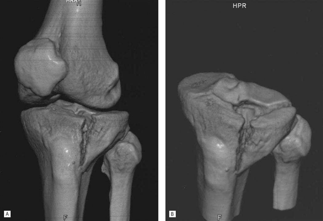
Fractures of the tibial plateau
Fractures of the tibial plateau (Fig. 25.1) involve the proximal tibial articular surface. Fracture patterns vary greatly. Sometimes associated soft tissue damage may be quite significant. In the elderly, often a low energy mechanism of injury produces such a fracture. The goals of treatment should be the attainment of an accurate reduction and if at all possible the institution of early ROM exercises. Delayed weight-bearing often is necessary to prevent the fracture site from collapsing.
< div class='tao-gold-member'>


Stay updated, free articles. Join our Telegram channel

Full access? Get Clinical Tree


