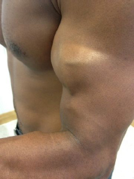
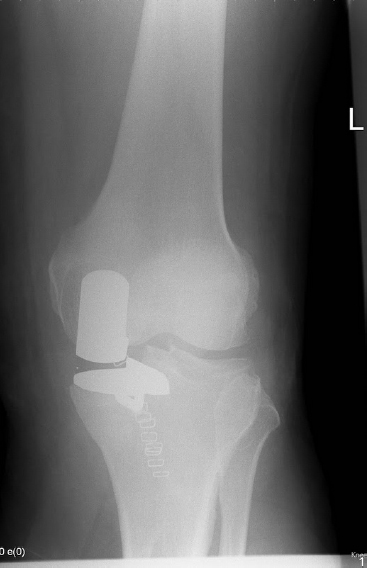
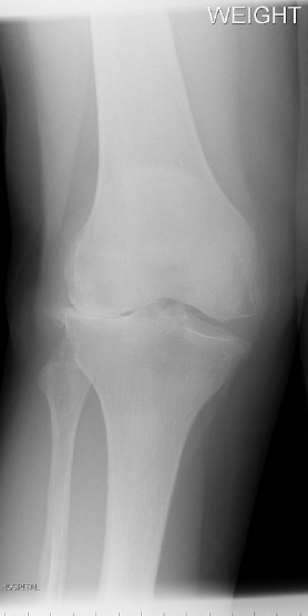
Adult Orthopaedics and Pathology 1. Clinical picture of tear of the distal biceps tendon. Please outline its causes and management? What is the prognosis of non operative and operative management? Please describe the principles of non operative and operative management and the relevant scientific evidence Causes Two theories 1. Reduced blood supply at zone two of tendon 2. Mechanical impingement in pronation – extension force in flexed elbow causes rupture Clinical features More common in men than women Middle age: during lifting or rotation of forearm Acutely, bruising antecubital fossa and tenderness 1. Reduced function – weakness particularly supination, flexion less marked 2. Loss of contour 3. Biceps squeeze test – less supination Management Imaging:Radiographs usually normal, ultrasound scan (operator dependent) and MRI Non operative -Acceptable outcome in less active patients – Cosmetic deformity is not an indication for surgery – cramping pain with prolonged use is rare -reduced flexion strength of 15 percentage and supination strength of 40 percentage Surgical A. Anatomic versus non anatomic Anatomic- re attach to radial tuberosity Non anatomic – Attach to brachialis (salvage procedure with chronic rupture, no increase in supination strength, can help with pain. Late reconstruction with hamstrings and palmaris longus have been described B. Two incision vs one incision Historically a single incision with drill holes and sutures was used, this carried a high risk to the Posterior Interosseus Nerve. Two incision approach of Boyd and Anderson was used giving less chance of nerve injury, but increased incidence of heterotrophic ossification and radio-ulnar synostosis The use of suture anchors has allowed a return to a single incision with reduced nerve risk with similar or superior outcomes reported. Ref: McKee et al JSES 2005 Describe radiograph- superior migration/glenoid erosion/Osteoarthritis Several options to classify this: Most straightforward FAVARD classification but more common HAMADA classification FAVARD: 1 to 3 1) Superior migration of the humeral head and changes visible on the inferior surface of the acromion 2) Central gleno-humeral space narrowing 3) Lysis of the humeral head or the acromion HAMADA Grade 1 to 5 Management -Arthroplasty (hemi/ reverse?) Pre operative factors 1. Cuff deficiency 2. Bone stock (assess with pre-operative CT scan) 3. Wear pattern 4. Soft tissue status Crucial concept compensation for tear: About 50% of patients with arthritis and massive cuff tear can elevate limb: acceptable outcome with lower risk with hemi or re-surfacing. 50% of patients are unable to elevate the limb with a cuff tear and arthritis. In these patients, consider reverse shoulder arthroplasty Principles of reverse shoulder arthroplasty -Medialises gleno humeral joint center of rotation- lengthen glenoid lever arm- increases power -Humeral head lowered-increases tension on deltoid- increases power Disadvantages with reverse Complication rates are high (up to 50% Werner et al) Limited survival and difficult to revise (5 -30% at 5 years) In Europe, reverse shoulder arthroplasty is used more commonly that in North America Problems associated with reverse shoulder arthroplasty 1. Instability- dislocation 2. Polyethylene wear 3. Lack of external rotation 4. Scapular notching 5. Are there long term results? Plenty! Good gains reported in pain and function reflected in Constant score and Oxford shoulder score Caution 1) Using reverse in acute trauma 2) Do not implant a total shoulder arthroplasty if the rotator cuff is torn, unless the tear can be repaired Rotator cuff tears Common pathology increase with age: approximately 30% has Partial thickness tear or full thickness tear at 70 years Genetic basis: siblings of patients with symptomatic tears more than twice as likely to have a tear and four times to have symptoms than controls Not all torn cuffs need to be repaired Investigations: Radiographs: in the axial view, beware of os acromiale Ultrasound scan good for tear identification, role in partial tears not proven MRI Tear size, retraction, repairability and fatty substitution (Goutallier 0-4, first described in CT, validated for MRI) 0 = normal, 1 = fatty streaks, 2 = more muscle than fat, 3= same muscle and fat, 4= more fat than muscle Higher grades associated with worse surgical outcome Management Conservative treatment: injection and physiotherapy Indications for cuff repair Symptomatic tears failing to respond to conservative treatment in appropriate patients. Significant intervention and rehabilitation prolonged (six weeks sling, six months to recover). Not without risk. 2. Proven tear – Ultrasound scan/MRI 3. Reduced activities of daily life and night pain Aim of surgery- reduce pain and improve function Methods Evidence suggests re-tear rates up to 50% Failed repairs can do well in outcome except on strength Principles of rotator cuff surgery Consider fusion if os acromiale Main techniques -Open -Arthroscopic subacromial decompression and mini open repair -Arthroscopic (A controversial area, with each having advantages and disadvantages) Acromioplasty (pain relief) Restoration of bone tendon interface (mechanical strength) Stimulation of healing (bone) Removal of scarred atrophic tendon edges Protection of repair (immobilisation) Restoration of range of movement and power (rehabilitation) Causes Uncommon but well recognised complication of fractures, particularly paediatric distal humeral fractures. Late presentation with ulnar nerve symptoms of variable severity. Associated with varus angulation Treatment options Transposition ulnar nerve Distal humeral osteotomy Choice of transposition: submuscular or subcutaneaus Surgeon preference rather than scientific evidence 4. Radiograph of elbow replacement. Principles and complications This are is an antero-posterior and lateral radiographic views of the elbow There is a: 1) Total elbow replacement (radial head usually excised) 2) A prosthetic radial head (usually for trauma) Much less common 3) A distal humeral replacement 4) Lateral column replacement Elbow replacements are performed for arthritis (inflammatory, bleeding disorders, osteoarthritis, and post traumatic) They may also, in certain circumstances, be performed acutely for trauma (pre-existing disease or complex fracture pattern intraatricular in low demand patient). The concept is reduction of pain and improvement in range of movement. There are numerous prostheses on the market They may be broadly classified as follows: -linked (more stable but greater stress concentration, so concerns about longevity) -un-linked (they rely on the integrity of muscles and ligaments, hence concerns about stability) Complications: The complications of any surgical procedure: In addition consider: 1. Increased infection in immunosurpressed rheumatoid patients 2. Wound problems (5%) 3. Deep infection 4. Ulnar nerve neurapraxia (usually transient) 5. Per-operative fracture (consider fixing or linked prosthesis) Winging scapula may result in severe shoulder dysfunction Causes 1-Trapezius palsy 2-Serratus anterior palsy 3-Sprengel deformity (not truly winging) 4-Fascio-scapular-humeral dystrophy (FSHD) 1. Trapezius Palsy Commonly a result of injury to the spinal accessory nerve (iatrogenic, can be intentional with neck clearance) Difficulty in abducting arm without pain Treatment: physiotherapy and expectant attitude If symptoms persist, consider EMG and neurolysis In chronic cases, consider muscle transfer (Eden –Lange procedure) 2. Serratus Anterior Palsy The long thoracic nerve (C5, C6, C7) is involved Causes: viral illness, recumbency, Pregnency, idiopathic Recovery after closed injury: usually 1 year, but it can take up to 3 years Treatment: physiotherapy Braces often poorly tolerated Surgery: transfer of sternal head of pectoralis major to the inferior pole scapula with fascia lata. 3. Sprengel’s deformity, the scapula is high and hypoplastic. It produces variable disability, and surgery is not usually needed It can be associated with other congenital abnormalities (eg. Klippel–Feil syndrome) 4. Facio-scapular-humeral dystrophy (FSHD) Autosomal dominant Unilateral presentation in teens, progressing to bilateral involvement Surgery: scapulothoracic fusion These are anteroposterior and lateral radiographs of the elbow showing loss of joint space, subchondral sclerosis and ostephyte formation. This appearance is in keeping with a diagnosis of osteoarthritis. Ostearthritis of the elbow may be primary (male manual workers) or secondary to inflammatory joint disease, trauma and infection The main presenting complaints are loss of range of movement and pain. Ulnar nerve symptoms and signs may also be present. Locking may be a feature. Examination, to include range of movement and distal neurology. Treatment tailored to symptoms Conservative: analgesia, physiotherapy and injection (corticosteroids, hyaluronic acid). Surgical options: 1. Arthroscopy: can be a simple washout, progressing to loose body removal, debridement and radial head excision. 2. Outerbridge –Kashiwagi (OK) procedure Posterior approach with joint debridement, loose body removal, and if needed ulnar nerve transposition (please see above re: safety and result of ulnar nerve transposition). 3. Radial head excision for isolated disease 4. Soft tissue releases to improve range of movement 5. Interposition arthroplasty may be an option in young patients as a salvage for post-traumatic osteoarthritis. 6. Elbow arthroplasty Mr Paul Allen Consultant Knee surgeon Princess Alexandra Hospital 1. Shown clinical picture of infected Total Knee Replacement How will you proceed HISTORY 1. Wound Infection 2. Post op drainage/antibiotics 3. Co morbidities- Diabetes/smoking/obese/steroids EXAMINATION 1. Swelling/warm/pain/reduced range of movement/Sinus Investigations 1. Bloods –White cell count /CRP/ESR 2. Scans –White cell scan 3. Aspirate (after stopping antibiotic for two weeks) – sent for gram stain and culture -if first aspirate negative and clinical features continue – send two more aspirates -Arthroscopic synovial tissue biopsy Classification 1. Positive intra-operative culture in revision 2. Early –less than four weeks–superficial/deep 3. Acute haematogenous 4. Late chronic-more than four weeks Treatment protocol 1. Early superficial -no arthrotomy -debride/irrigate -Close wound -Antibiotics for six weeks 2. Early deep -change polyethylene -debride/irrigate -Close/Antibiotic beads medial and lat gutter -Antibiotics for six weeks 3. Acute haematogenous –less than four weeks – same as early deep 4. Chronic- late more than four weeks Options are -Two stage revision (gold standard) -One stage revision Two stage revision First stage -Remove all components and thorough debridement -Antibiotic cement and spacer -Antibiotics for six weeks or more – Repeat blood markers/CRP -Wait two weeks after CRP return to normal/clinically normal -Second stage revision /re implant after six weeks or more Single stage revision option if organism known before hand/patient cannot withstand two stage 2. Shown MRI of knee with osteochondral defect. How will you manage Definition – Avasclar necrosis occurring at osteochondral junction in second decade males most commonly around knee (also ankle/elbow) Most common site – lateral aspect of medial femoral condyle Etiology 1. Repetetive micro trauma 2. Genetic 3. Altered micro vascularity Clinical features 1. Pain/ crepitus/loose body/Quadriceps wasting/effusion 2. Wilson test –flex knee 90 degree and internally rotate- extend- pain at 30 degrees is Wilsons test positive Investigations 1. Radiograph – tunnel view 2. MRI Classification Guhl arthroscopic 1. Softening of cartilage 2. Undisplaced 3. Displaced but attached 4. Loose body Treatment Depend on 1. Age- juvenile heal better 2. Size 3. Area- weight bearing 4. Guhl stage Non operative 1. Juvenile (less than 12 years) OCD, painless or painful with non displaced lesion-Reduced weight bearing plus quadriceps strengthen for 3-6 months/follow up with MRI to assess healing Indications 1. Failed non operative in skeletally immature individuals or adults 2. Unstable/loose body 3. Avascular segment 4. Weight bearing area with large lesion Goals 1. Pain relief/prevent osteoarthritis/better function/promote healing Treatment options 1. Arthroscopic drilling 2. Curettage+ drill+Bone graft if big defect 3. Unstable segment –screw fixation after curettage and drilling 4. Osteo chondral allografts /autografts – Mosaic plasty (recommend by NICE with special consent/audit/research.IPG162-march 2006) -Autologous chondrocyte implantation(ACI) (not recommended by NICE .TA89-issued may 2005-for review in 2013) 3. UNICONDYLAR KNEE REPLACEMENT. Radiograph of unicondylar knee replacement. Discuss merits and demerits Types Mobile/fixed bearing Advantages over Total Knee Replacement 1. Less complication 2. Less time/blood loss/dissection 3. More Range of movement 4. Better patient satisfaction 5. Easier surgery/cheaper 6. Early rehabilitation/return to work Disadvantages 1. 90% 10 year survival – less than TKR 2. Feasible only if one compartment involved 3. Needs good range of movement with varus or valgus malalignment less than 10 degrees 4. 10 % revision rate compared to 3% for total knee Radiograph of valgus knee with osteoarthritis. How will you manage. What will you do different from a routine knee replacement? Examine 1. Correctable deformity? 2. Fixed flexion deformity? 3. Valgus or varus malalignment 4. Common peroneal nerve neurology 5. Patella tracking Evaluation 1. Radiographs -Weight bearing AP-lateral in 30 degree/sunrise, skyline views/Pelvis -Ascertain the presence of osseous defects, and quantify their extent -Assess amount of bone resection-template is a must Assess severity of valgus 1. Mild –less than 10 degree
Related posts:


Stay updated, free articles. Join our Telegram channel

Full access? Get Clinical Tree


