Open Reduction and Internal Fixation of Tibial Plateau Fractures
Introduction
Tibial plateau fractures involve joint surface, metaphysis of proximal tibia
Occur from high-energy and low-energy mechanisms
Consider severity of injury to bone and surrounding soft tissues when determining management
Goals—Anatomic reduction, restoration of mechanical axis, stable fixation enabling early range of motion (ROM), preservation of surrounding soft tissues, avoidance of infection, ligamentous repair, or reconstruction as needed
Patient Selection
Careful examination of injured extremity is mandatory
Vascular injury, compartment syndrome, and open injuries should be treated emergently
Note knee stability and soft-tissue conditions and image appropriately
Indications
Absolute, for open treatment—Open fractures, compartment syndrome, vascular injury
Relative—Joint instability or malalignment, medial condylar fractures, lateral plateau fractures with displacement greater than 3 mm, condylar widening greater than 5 mm, patient with multiple traumatic injuries
Consider closed treatment for fractures with less than 2 mm of joint surface displacement, normal alignment, and stable knee in extension
Contraindications
Consider existing comorbidities, baseline functional level
Delay open treatment until surrounding soft tissues have time to heal, when possible
Goals—Maintain viability of soft tissues, preserve vascularity surrounding fracture
Alternative Treatments
For bicondylar fractures—Alternative technique uses small-wire ringed external fixator
For minimally displaced lateral split fractures with minimal joint surface comminution—Closed reduction with percutaneous fixation is an option; rule out lateral meniscus injury or incarceration preoperatively with MRI
For some fractures with intact or restorable cortical envelope—Arthroscopically assisted reduction and fixation
Classification
Two classification systems describe fracture pattern but do not consider ligamentous or soft-tissue injury or predict outcomes
Schatzker classification
Six types
Types I, II, III—Low-energy mechanism; involve lateral plateau
Types IV, V, VI—Higher-energy mechanism; involve medial plateau or bicondylar injury
AO/Orthopaedic Trauma Association classification
Classified as extra-articular, partial articular, and complete articular
Further subdivisions based on fracture severity
Preoperative Imaging
Radiography
AP, lateral, and internal/external oblique views of knee
AP with beam directed 10° caudal shows articular surface displacement
Computed Tomography

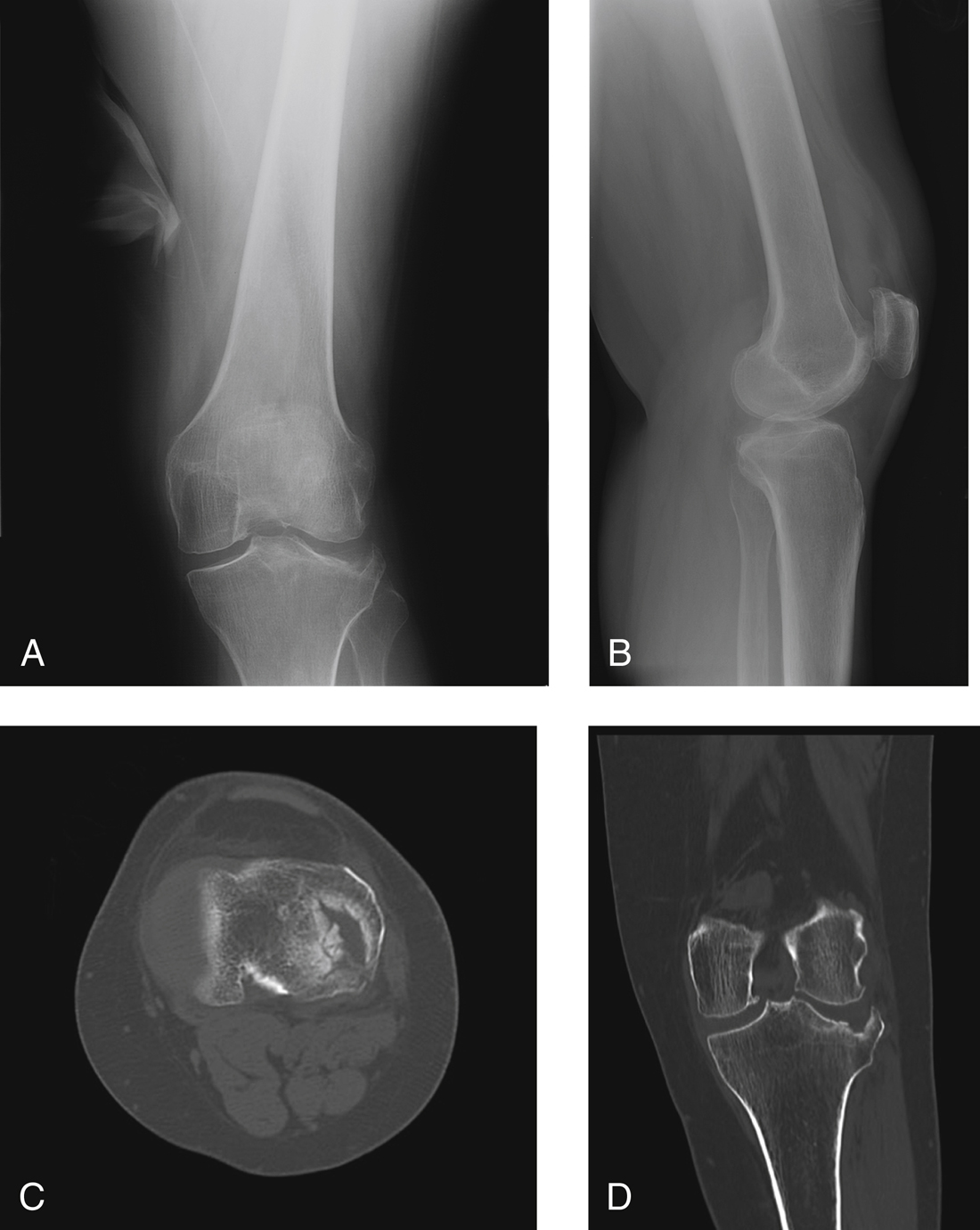
Figure 1AP (A) and lateral (B) radiographs show a Schatzker type III tibial plateau fracture. Axial (C) and coronal (D) CT cuts demonstrate a severe articular depression with no associated split in the cortex.
Axial with sagittal, coronal reconstructions useful in surgical planning
Helps evaluate articular depression (Figure 1)
Magnetic Resonance Imaging
Preoperative MRI helps evaluate for soft-tissue injury; meniscal, ligamentous injuries common
Helps evaluate need for ligamentous repair before hardware placement
| Video 76.1 Open Reduction and Internal Fixation of Tibial Plateau Fractures. Mark E. Nake, MD; James A. Goulet, MD (21 min) |
Procedure
Approaches
Anterolateral approach—For most common fractures (Schatzker types I, II, and III) and fixation of lateral plateau with dual-incision technique in bicondylar fractures
Lobenhoffer posteromedial approach—For fixation of medial plateau
Direct posterior approach—Rarely used; for posterior shear–type injuries
Stay updated, free articles. Join our Telegram channel

Full access? Get Clinical Tree


