Open Reduction and Internal Fixation of the Tibial Plafond
Patient Selection
Articular surface or gross metadiaphyseal malalignment of tibial plafond requires open reduction and internal fixation
Intrinsic articular surface impaction occurs medially on plafond, from internal rotation/supination, or laterally, from external rotation/pronation from axial load
Fibular involvement common; Volkmann fragment with or without ligamentous avulsion possible
Contraindications to surgery include noncompliance with cessation of nicotine use or intravenous drug addictions, venous insufficiency, and systemic disease; evaluate on case-by-case basis
Preoperative Imaging

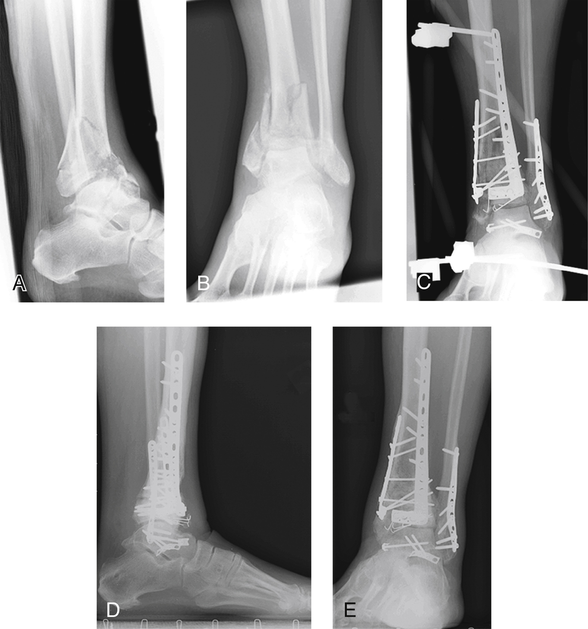
Figure 1Radiographs show initial injury, treatment, and outcome of a tibial plafond injury. Lateral (A) and mortise (B) radiographs show the injured ankle and foot. C, Mortise postoperative radiograph shows a medial buttress plate. The frame was maintained in place for soft-tissue management. Lateral (D) and mortise (E) radiographs at 1.5-year follow-up.
Plain AP, lateral, and mortise radiographs of ankle (Figure 1)
For tibial plafond fracture, obtain imaging throughout treatment course
Procedure
Room Setup/Patient Positioning
Use similar surgical setup for index, definitive surgeries
Supine position on pressure-relieving mattress on radiolucent table
Place wedge under ipsilateral hip to allow external and internal rotation of ankle
Index surgery uses more internally rotated position; definitive surgery uses internal and external rotation
Place ramp pad under ipsilateral limb; allows access for surgery, intraoperative radiographs
Surgical Technique
Two surgeries are usually necessary
Index surgery provides gross reduction of ankle through distraction, with talus under tibia, via external fixator
Obtain CT after external fixation to plan definitive intervention
Definitive surgery occurs 10 to 21 days after injury
Index Surgery

Figure 2Photographs show an external fixation device. A, A unicolumnar frame is shown after definitive surgery. The two tibial pins are placed outside the affected area and as far apart as possible. Note the incisional wounds associated with the fibular fixation and the anterolateral approach. B, A typical bicolumnar frame is shown. When a bicolumnar external fixation device is needed, the standard arrangement includes two bars to tie the two tibial pins to the medial calcaneus and midfoot (cuneiforms). This arrangement includes the components used in a unicolumnar medial application (A), but the calcaneal pin exits the lateral side, and a third bar ties it to the proximal tibial pin (B). In the arrangement shown, the midfoot pin through the cuneiforms was not needed to provide adequate stability.

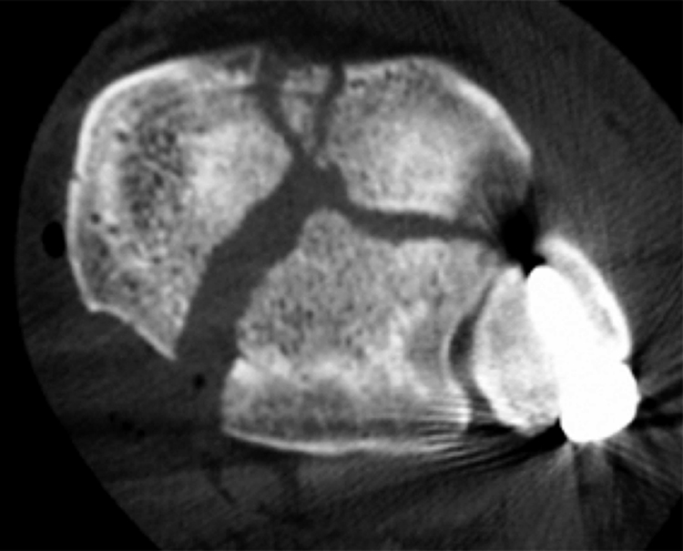
Figure 3CT scan shows the three main fragments in a tibial plafond injury: the posterior or Volkmann fragment that is attached to the fibula via the tibiofibular ligament; the medial fragment, which in this case includes the malleolus; and the anterolateral or Chaput fragment.
Occurs within hours of presentation
Early external fixation enables earlier definitive treatment, improved outcomes
Place Schanz pins carefully to avoid neural and vascular structures
Address fibular injury first
Must restore correct fibular length, orientation
Fibular reduction more critical with associated tibial plafond injuries; posterior (Volkmann) fragment connected to fibula via posterior tibiofibular ligament
Unless ligament avulsed, fibular reduction grossly reduces Volkmann fragment; anatomic fibular reduction important for good outcome
Fibular fixation can wait until trained personnel and the needed equipment are available
Place external fixator after treating fibula
Place basic frame along medial column (Figure 2, A); lateral injuries may need bicolumnar frame (Figure 2, B)
Attach medial column frame to tibia, calcaneus, and medial cuneiform
Locate all pins outside anticipated location of incisions
Insert calcaneal pin first
Terminally threaded, inserted medially for medial column frames; centrally threaded for bicolumnar frames
No predrilling required
Place in posterior portion of calcaneal tuberosity, posterior to line connecting superior, posterior edges; avoids sural nerve, calcaneal branch of tibial nerve
Insert proximal tibial pin
Place at proximal middle third junction of tibia through anteromedial surface; avoid saphenous nerve and vein
Use 5-mm Schanz half pin
Predrilling required
Insert third pin through medial, middle, lateral cuneiforms
Insert through medial surface of medial cuneiform
No predrilling required
Place distal tibial pin last; location determined by tibial fracture; place as distally as possible, while remaining outside fracture area, forthcoming internal fixation
Stay updated, free articles. Join our Telegram channel

Full access? Get Clinical Tree


