Open Reduction and Internal Fixation of the Distal Radius With a Volar Locking Plate
Patient Selection
Surgery involves closed or open reduction and internal fixation
An alternative is closed reduction (or repeat closed reduction)
Preoperative Imaging
Plain Radiography

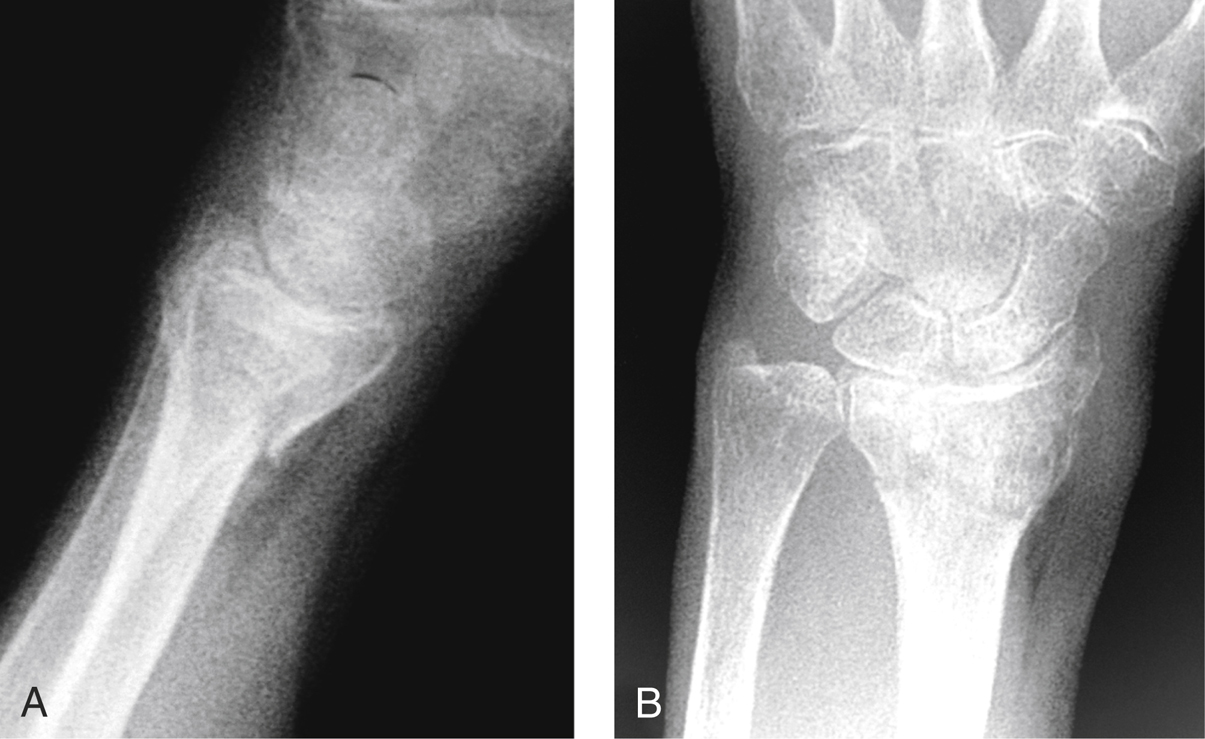
Figure 1Lateral (A) and PA (B) image-intensifier views show a fracture of the distal radius.
PA and lateral radiographs before and after manipulative reduction (Figure 1)
Traction radiographs may be helpful
Computed Tomography
Not always indicated, but for certain fracture patterns they may provide further details about number, size, location, and displacement of articular fractures
Three-dimensional reconstructions are easy to interpret
Procedure
Surgical Technique

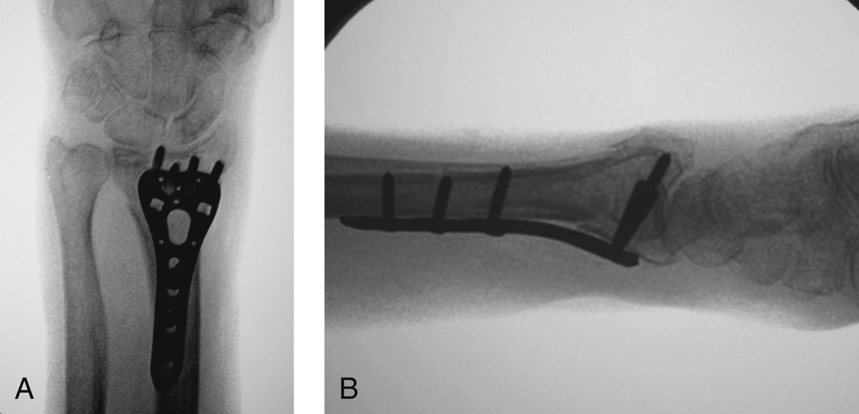
Figure 2Intraoperative PA (A) and lateral (B) image-intensifier views show plate position and fracture alignment.

Stay updated, free articles. Join our Telegram channel

Full access? Get Clinical Tree


