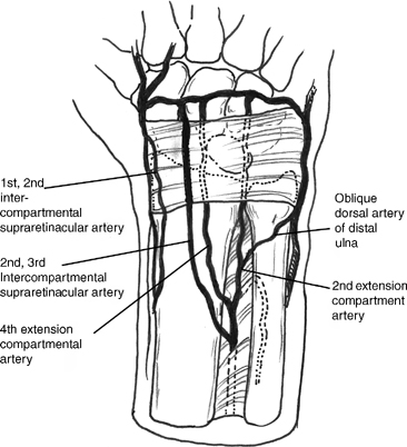
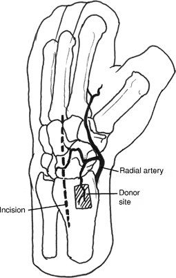
31 Open Reduction and Internal Fixation of Scaphoid Nonunion with Vascularized Bone Graft Figure 31-1 Figure 31-2
Indications
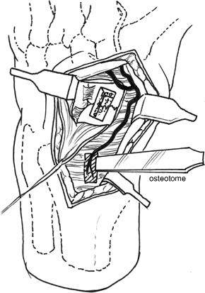
Technique
Related posts:
 Intra-articular Fractures of the Distal Radius Treated with Dorsal Plate
 Resect Ulnar Styloid Fracture with Repair of Triangular Fibrocartilage Complex
 Sauve-Kapandji Procedure
 Flexor Carpi Radialis Tendon Stabilization of the Scapholunate Joint (Brunelli Procedure)
 Closed Reduction and Internal Fixation of Bennett’s or Rolando’s Fractures
 Capitate Shortening with Capitohamate Fusion
Stay updated, free articles. Join our Telegram channel

Full access? Get Clinical Tree


