Open Reduction and Internal Fixation of Scaphoid Fractures
Patient Selection
Scaphoid fractures represent 11% of all hand fractures; occur mainly in men between the ages of 20 and 30 years
Unrecognized injury and/or inadequate treatment can result in nonunion, which can lead to chronic pain, functional loss, and arthritis
Indications
Displacement
Carpal collapse
Comminution
Desire to avoid prolonged immobilization
Risk factors for delayed union, including delay to diagnosis, proximal fracture type, or other patient factors
Contraindications
Comorbidities (relative)
Inability to tolerate anesthesia (relative)
Preoperative Imaging
Radiography

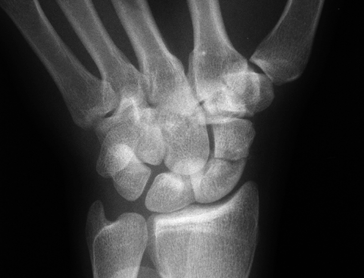
Figure 1Scaphoid view radiograph, taken with the wrist in extension and ulnar deviation, demonstrates a scaphoid waist fracture.
PA, lateral, and oblique and navicular view radiographs of wrist; look for loss of carpal height, intercarpal widening, carpal malalignment, associated fractures
Navicular view (30° extension with ulnar deviation) demonstrates long axis of scaphoid (Figure 1)
Nondisplaced fractures may not appear on initial radiographs
Bone Scan
Technetium methylene diphosphonate (MDP)-99 has high sensitivity and specificity
Must wait 48 to 72 hours after injury
Mostly of historical interest
Magnetic Resonance Imaging

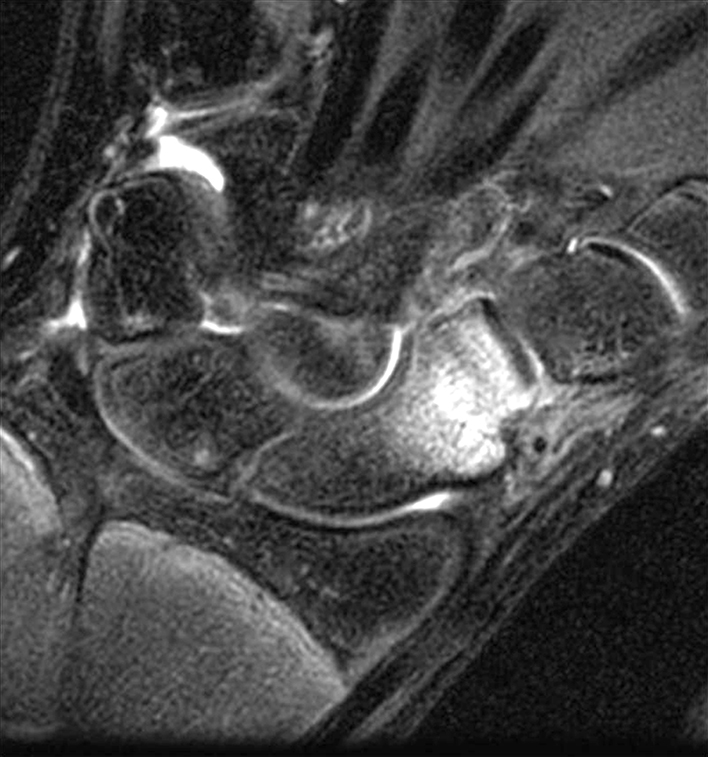
Figure 2A coronal T2-weighted MRI of the wrist demonstrates increased signal intensity of the distal portion of the scaphoid, consistent with an occult fracture.
High sensitivity and specificity reported (Figure 2)
May perform as early as 24 hours after injury
False-positive results exist because of marrow edema
CT Scanning
May be used for diagnostic purposes or for preoperative planning
More commonly used for evaluation and treatment planning in the chronic setting to evaluate for bone loss or bone healing than in the acute setting for diagnosis
Preferred Evaluation Sequence
Radiographic series
If fracture not demonstrated but high suspicion exists, consider immobilization in a thumb spica cast
High-demand patients may undergo MRI
Low-demand patients or those who prefer watchful waiting may have repeat radiographs and clinical examination after 10 to 14 days of immobilization
Nondisplaced fractures particularly of the distal scaphoid or even of the waist, or those that are visible only on MRI, are most commonly amenable to nonsurgical treatment; surgical treatment may be chosen to allow return to activity sooner
Procedure
| Video 43.1 Open Reduction and Internal Fixation of Scaphoid Fractures. Kristofer S. Matullo, MD; Alexander Y. Shin, MD (2 min) |
Room Setup/Patient Positioning
Supine position with radiolucent hand table
Obtain preoperative C-arm fluoroscopy images to guarantee visualization
Apply tourniquet around upper extremity
Use regional block or general anesthesia
Consider preoperatively if bone graft will be required; consider obtaining consent and positioning should iliac crest, olecranon, or distal radius bone grafting be needed
Special Instruments/Equipment/Implants
C-arm (mini or standard)
Cannulated headless compression screws
Surgical Technique for Volar Approach

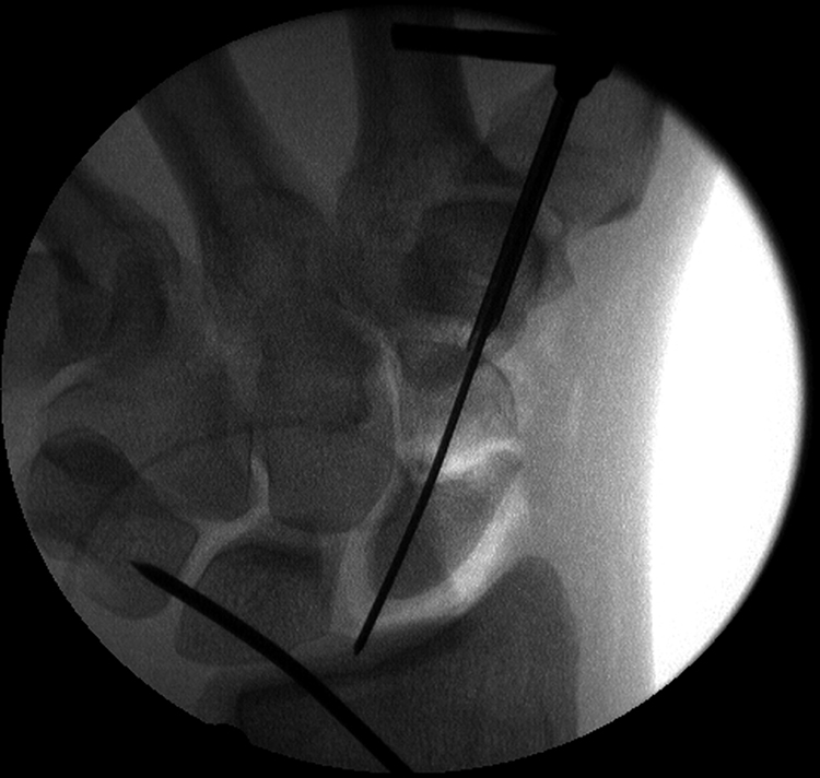
Figure 3Intraoperative fluoroscopic image demonstrates the guidewire along the central axis of the scaphoid.

Stay updated, free articles. Join our Telegram channel

Full access? Get Clinical Tree


