Image 8.1
MRI sagittal T2 view of a complete triceps tendon lesion
8.3.7 Triceps Snapping
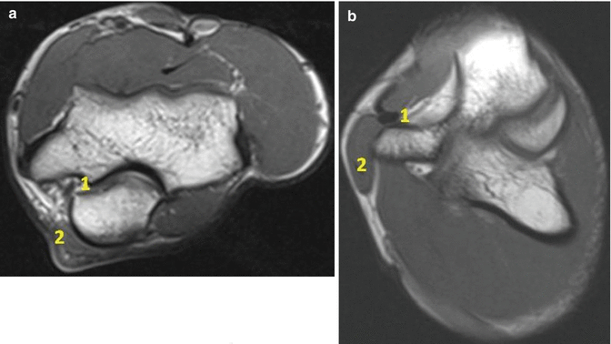
The clinical study of the triceps snapping is usually performed with the patient’s elbow on the examiner’s open hand, with the thumb over the medial epicondyle; a passive movement from extension into flexion can reveal a single or double snap with anterior direction; if a double snap is found, the ulnar nerve determine the first one, followed by the medial head of the biceps [59]. The imaging study of triceps snapping can benefit of ultrasounds, most of all with a direct dynamic evaluation [60] of the ulnar nerve and/or of the medial head of the triceps; the MRI finds its utility, if the study is performed with the elbow in flexion [61] (Image 8.2).


Image 8.2
MRI coronal T1 view of a case of triceps snapping: (a) in full extension, the snapping is not demonstrable; (b) in 120° of flexion, the MRI demonstrates a triceps snapping and dislocating ulnar nerve
8.3.8 Olecranon Bursitis
The diagnosis is clinical, based on a conspicuous swelling on the posterior elbow, caused by the effusion that develops because of bursal inflammation. Dealing with pain, in septic bursitis, all the elbow movements are painful, while in aseptic bursitis, pain is low or absent. The features of aseptic and septic olecranon bursitis, if compared, do not show significant differences.
The skin temperature over the bursitis is higher in septic cases and normal in aseptic. Ultrasound scanning can be used for differential diagnosis in elbow swelling (synovial proliferations, calcifications, loose bodies, rheumatoid nodules, gouty tophi) and to evaluate the general quality of the fluid. Standard X-ray of the affected elbow is useful to exclude bony lesions and to study the joint. Rare is the use of MRI [50].
8.4 Treatments
8.4.1 Valgus Extension Overload Syndrome
The first treatment is conservative: rest from sport, NSAIDs, ice, and rehabilitation therapies, at least for the first 4 weeks. From the fifth week, a program of strengthening of active elbow stabilizers, the full elbow movement recovery, and a plyometric rehabilitation can be started. The restart of the sport-specific gesture with a specific and progressive program can be planned between the 16th and the 24th week from the beginning of rest.
Kancherla suggests rest and pitching limitation for 2–6 weeks, followed by a sport-specific rehabilitation (dynamic stabilization, reinforcement of flexor/pronator mass, in particular with eccentric exercises) and an interval throwing program; if this program is not effective in pain resolution, this author suggests to evaluate for surgical treatment [21].
An imaging evidence of posteromedial spurs should drive the choice toward surgical removal. Arthroscopy is very useful in this disorder, because it gives the possibility to assess the anterior compartment, looking for chondral lesions, loose bodies, and instability signs (by direct view of UCL anterior bundle or by the indirect sign of the medial joint side opening of 1 mm at 70° of flexion) [62].
Cohen underlined that arthroscopic debridement, olecranon spur excision, and loose body removal allow return to throwing sports and reliable subjective and objective results in carefully selected patients [52].
The posterior compartment study gives the possibility to treat olecranon spurs and analyze olecranon fossa. In literature, it has been proved that an olecranon resection lower than 8 mm. is not dangerous for an iatrogenic loss of ulnohumeral constraint and a consequential strain increasing on UCL, but we consider prudent to remove just the spurs and the posterior scar. After an arthroscopic arthrolysis, the rehabilitation program can be started after 2 weeks with the aim of coming back to the field between 3 and 6 months.
8.4.2 Olecranon Stress Fracture
Both conservative and surgical modalities have been purposed for the treatment of olecranon stress fractures. Several authors refer good results with conservative treatment based on rest, splint, and progressive return to sport [2, 42, 53]. Surgical treatment, however, is considered by some authors the better option for these patients, primarily with the aim of a quick competitive sport return [7] and secondly because the incidence of nonunion and delayed union is higher after conservative treatment and requires secondary intervention [7, 63, 64].
Lu suggests that minimally or nondisplaced transverse fractures respond successfully to conservative measures, including activity restriction or immobilization with splint/cast [38]. For those with displacement greater than 2 mm, surgical treatment leads to good results and lower nonunion rates. Lu also agree with Suzuki’s statements [7] suggesting an early surgical approach for oblique olecranon stress fractures. Symptomatic tip fragments should be excised [2, 65].
Orava proposed the use of tension band for transverse fractures and screw in compression for the oblique ones [6]. Furthermore, arthroscopically assisted procedures can allow for additional diagnosis of associated lesions (loose bodies, osteophytes, ligament injury, and chondral damage). The postoperative treatment for olecranon stress fractures is based on splint with 90° of flexion for 7–10 days, followed by a 4-week rehabilitation with passive and then active flexion and extension and active pronation and supination, full active movement allowed at the sixth week, strengthening exercises during the eighth week, and interval throwing program in the 12th week.
8.4.3 Persistence of the Olecranon Physis
As for the previously described disorders, the initial management consists of rest, cessation of throwing, nonsteroidal anti-inflammatory drugs (NSAIDs) and ice, that can be successful in most patients. Surgical treatment may be beneficial after failing conservative management for 3 or 4 months, preferring low-profile systems, as tension-band wire construct and a single lag screw have been described as successful fixation options [35, 66]. Arthroscopy is not routinely used but can be useful in the cases of associated chondral lesions, that need a treatment.
After the surgery, a removable splint is usually used for 3 weeks, with passive and active movement from the second week; interval throwing program is allowed after 8 weeks and return to competition usually needs 4 months.
8.4.4 “Boxer’s Elbow”
The treatment of boxer’s elbow is based, firstly, on nonsurgical treatment (rest, ice, compression, elevation), physical therapy, NSAIDs, and eventually corticosteroid injections of the posterior side of the elbow. If this approach fails, surgical treatment can be considered. Elbow arthroscopy with debridement of the olecranon is usually the first choice, because it allows scar tissue and loose bodies removal, resection of posterolateral osteophytes, and partial resection of the olecranon tip. An arthroscopic stress test, to evaluate the medial opening, should always be performed [44].
8.4.5 “Handball Goalie’s Elbow”
Basing on similar pathogenesis and clinical onset, handball goalie’s elbow is treated in the same manner as VEOS.
8.4.6 Triceps Tendon Lesions/Tendonitis
Historically, partial tears of the triceps have been treated conservatively [57, 67, 68]. In his work on professional American football players, Mair [69] evaluated 11 complete ruptures and 10 partial ruptures; the author underlines that the extent of the tear may help to decide whether early surgery is necessary: MRI lesions of 90–100 % of the triceps tendon should be treated with early surgical repair. Partial tears (involving 75 % of the tendon on MRI or less) show the capacity to heal in some instances.
According to Morrey, partial tears can be treated nonoperatively for 6–8 weeks: if the symptoms does not disappear after this period, the patient should be surgically treated; for complete ruptures, immediate surgery is the treatment of choice. If the lesion is treated acutely, a direct suture of the tendon to the olecranon with nonabsorbable sutures is indicated. If a delayed reconstruction must be performed, Morrey suggest the anconeus slide, in the cases with minor defects and if this muscle is intact, or the Achilles tendon allograft for major lesions [49].
8.4.7 Triceps Snapping
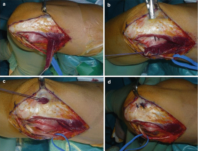
Triceps snapping in athletes, when the symptoms are not tolerated and negatively affect the performance, can be surgically treated: a release of the medial head of the triceps is followed by a reattachment in a more lateral position, so to avoid its snapping over medial epicondyle. In some cases, a simple removal of the medial head of the triceps can be performed without compromising the triceps strength. Ulnar nerve decompression is usually performed, its anterior transposition is evaluated basing on the symptoms and on the specific findings on the surgical field (Image 8.3).


Image 8.3
Surgical images of the triceps snapping case, reported in the MRI of Fig. 2: (a) portion of the medial head of the triceps detached; (b) preparation for transposition; (c) portion of the medial head of the triceps transposed in a more lateral position; (d) suture of the portion of the medial head of the triceps into the new position with clinical disappearance of snapping
8.4.8 Olecranon Bursitis
Conservative management is indicated, as the first step, in non-painful cases: the patient should be advised to avoid repetitive movements and the elbow should be protected with a bandage or, in cases of major swelling, with a brace. Comorbidities have to be treated with specific therapies.
In painful conditions or in the cases where the suspect of infection is high, a liquid aspiration from bursitis should be performed in aseptic conditions, for microbiological study, white cell counts, and glucose level quantification; a concomitant blood sample is desirable, because it is useful to compare the amount of white cells and glucose in the two samples: a fluid glucose level of less than 50 % of the serum level is suggestive for infection. Steroid injection into the bursa must be carefully evaluated, because of the high complication rate (infections, skin atrophy, chronic pain), that with aspiration alone are absent. After the aspiration, a compressive bandage is needed. We prefer to use a splint with 90° of flexion for 5–7 days, with the aim to aid soft tissue healing, avoiding the movement.
Aseptic olecranon bursitis that cannot find a solution with these treatments and septic bursitis need surgical excision; in the septic cases, specific antibiotic therapy must be extended as necessary [50].
8.5 Pearls of the Treatment/Prevention
Based on biomechanical and epidemiological considerations, the posterior elbow pain can be determined from a set of traumatic factors, that can create several lesions over the whole elbow. Once defined the surgical indication in the athlete with olecranon posterior pain, Paci suggests that the surgeon keeps a prudential attitude, treating the whole pattern of elbow lesions (loose bodies removal, spurs resections, UCL reconstruction), considering that these lesions find a common pathogenesis, with the aim to recreate a correct biomechanics, that is the base for a maximum sport-oriented recovery [39].
The key to success with VEOS and “handball goalie’s elbow” is the early recognition of the condition and the careful conservative management of the symptoms with appropriate periods of rest. If those conservative measures fail, arthroscopic surgical management is typically successful in returning the athlete to competitive sports at every level. Modification to throwing biomechanics may not necessarily improve clinical outcomes because the stresses from repetitive throwing may be the driving force to injury.
Olecranon stress fractures must be correctly diagnosed, classified, and treated, keeping in mind that a conservative treatment can be successful but that in high-level athletes, an aggressive approach can accelerate the return to sport and prevent delayed union and nonunion.
Persistence of the olecranon physis is treated without surgery in the majority of cases; if surgery is required, after the failure of conservative treatment, a synthesis with low-profile systems should be preferred; some authors suggest the use of bone graft to improve healing.
The boxer’s elbow is rare and can find its solution with conservative treatment, but often, arthroscopy of the posterior elbow is useful to obtain a quick sport return.
The approach for the posterior elbow pain in patients with skeletal immaturity must keep in highest importance the prevention [70] that needs a multidisciplinary approach (pediatrics, sports medicine, orthopedics, physiotherapy, etc.), aiming to preserve joint integrity and function and conciliating a healthy and harmonious growth with the sport. The prevention is the keystone of the athlete’s treatment, most of all for the younger ones, as well established with Baseball Little League for pitchers.
The surgery must be the last step of an articulated treatment and should be performed in the ideal physical and mental conditions (end of season, high motivation in return, etc.); the medical team must take care of every athlete “like a professional athlete,” well defining from the beginning all the steps of the treatment.
8.6 Results after Treatment (Evidence Based)
8.6.1 Valgus Extension Overload Syndrome
Reddy [71] refers a return to competitive sport after arthroscopy treatment of VEOS for 85 % of the athletes treated.
In case of medial instability, the arthroscopic treatment should be followed by a UCL reconstruction that can warrant a return to competitive sport in a percentage of cases from 81 to 95 in literature [72–74]. After a UCL reconstruction, the mean time to come back in competitive sport is at least 12 months. Dugas refers that the recurrence of the symptoms or clinical findings of VEOS are rare and have not been reported in any of the large series of elbow procedures in athletes. In his clinical experience, recurrence of posteromedial impingement is secondary to an underappreciation of the underlying medial ligamentous laxity and other predisposing pathology [27].
8.6.2 Olecranon Stress Fracture
Several authors refer good results with conservative treatment: Nuber suggests a treatment based on rest, splint, and progressive return to sport with good results [2]; Schickendantz refers that seven professional athletes with olecranon stress fractures came back to competitive sport with a personalized conservative treatment [42]. Patel expresses good results with rest, pitching avoidance, and limitation to complete extension for 4 weeks with splints; after this period, he allows full ROM and resistance, limiting the valgus stresses for 6 weeks; from the sixth week, sport-specific exercises are begun and from the eighth week, the interval throwing program can start [53].
Suzuki [7] and Nakaji [37], based on the good results obtained in their cases, suggest an early ORIF for olecranon stress fractures in the athletes. Paci performed an ORIF with a compression screw in 18 high-level athletes (in addition, two patients underwent a medial compartment reconstruction), that had poor results after conservative treatment, with a mean FU of 6.2 years. All cases showed the fracture healing and 94 % of patients return to the same or higher level of competition in a mean of 28 weeks. Despite the percentage of sport return and the good functional results, this study shows a high rate of concomitant surgical procedures and additional procedures: 6 of the 18 patients underwent hardware removal (two because of infection), two needed a second time reconstruction of the medial compartment because of persistent instability, and two patients needed olecranon spurs or loose bodies removal for unresolved pain. In the case series described by Paci, the return to competitive sport has been reached in a mean of 29 weeks (8–45) [39].
8.6.3 Persistence of the Olecranon Physis
As underlined by Charlton, the conservative treatment is successful in most patients [35]. However, resolution of symptoms can take as long as 4 months [75].
Dealing with surgery, the highest rates of successful union have been shown in patients undergoing bone grafting [35, 75], with the aim of filling the bone gap that this patients usually present. Charlton and Chandler found that operative stabilization with internal fixation and autogenous iliac crest bone grafting can resolve symptoms and allow a skeletally mature overhead athlete to return to previous throwing performance, maintained to a 32 months FU. Fixation alone, however, may lead to a 66 % failure rate [35].
8.6.4 “Boxer’s Elbow”
In [Valkering] case series, the arthroscopic treatment of five professional boxers with partial resection of the olecranon tip and removal of scar tissue and loose bodies brought to an improvement in ROM and a return to their preexisting level of boxing activity [44].
Stay updated, free articles. Join our Telegram channel

Full access? Get Clinical Tree


