Fig. 3.1
Cardiovascular disease and other major causes of death for all males and females in the United States in 2006 (Source for data: NCHS and NHLBI. Reproduced with permission from Lloyd-Jones et al. [1])
Recently, the prevalence of coronary heart disease (CHD) was shown to be 7.9 % in the United States (US) among adults age 20 years and older (Fig. 3.2) [1].
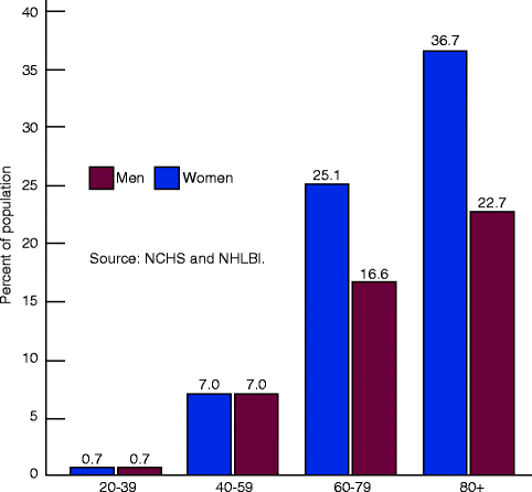
Fig. 3.2
Prevalence of coronary heart disease stratified by age and sex according to the National Health and Nutrition Annual Survey 2003–2006 (Reproduced with permission from Lloyd-Jones et al. [1])
Standard cardiovascular risk factors have been well validated in the prediction of coronary artery disease events. For example, increased levels of circulating low-density lipoprotein (LDL) cholesterol have been associated with increased CAD events particularly when present with other standard coronary artery disease risk factors such as increased age, male gender, active cigarette smoking, diabetes mellitus, and hypertension. The presence and severity of standard cardiovascular risk factors is the most important consideration in applying current cholesterol management primary prevention guidelines [2].
Risk scoring models, such as the Framingham Risk Score (FRS), have well-described limitations. For example, in the FRS, LDL cholesterol levels and family history of premature coronary artery disease are not directly included and the risk prediction provided is limited to 10 years. Furthermore, current prevention guidelines that are largely based on the FRS have never been prospectively proven to reduce mortality from cardiovascular causes [3]:
Among 222 relatively young patients (men <55; women <65 years-old) presenting with their first acute myocardial infarction, only 25 % would have previously qualified for preventative pharmacotherapy under the National Cholesterol Education Panel III guidelines prior to their cardiovascular event (Table 3.1) [2, 4].
Table 3.1
Mean (± standard deviation) cholesterol values among 222 young (men <55; women <65 years-old) patients presenting with their first acute myocardial infarction
Study population (mg/dL)
NCEP criteria (mg/dL)
Total cholesterol
190.1 ± 42.7
<200
LDL cholesterol
125.7 ± 38.6
<130
HDL cholesterol
Men
41.7 ± 14.9
>40
Women
45.2 ± 14.2
Triglycerides
145.0 ± 77
<150
Of 136,905 patients hospitalized with CAD, those patients without prior history of known CAD, other atherosclerotic vascular disease, or diabetes: 41.5 % had LDL < 100 mg/dL and 12.5 % had LDL < 70 mg/dL. Only 29.2 % of the patients without prior history of atherosclerosis or diabetes had an LDL > 130 mg/dL (Fig. 3.3) [5].
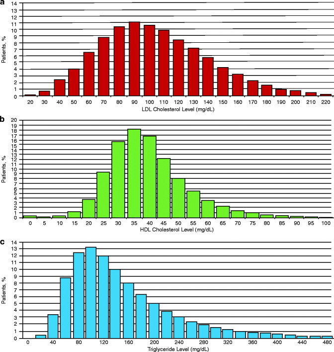
Fig. 3.3
(a–c) Distribution of admission cholesterol levels (LDL, HDL and triglycerides). (a) Histogram of admission LDL levels in 10 mg/dL increments. (b) Admission HDL levels in 5 mg/dL increments. (c) Admission triglyceride levels in 20 mg/dL increments (Reproduced with permission of Elsevier from Sachdeva et al. [5])
It is important to note that coronary atherosclerosis is an inflammatory process. Chronically increased degrees of inflammation are directly correlated to increased risk of plaque instability and resultant acute coronary syndrome (ACS) [6].
Evidence of coronary artery atherosclerosis and inflammation has been documented in infancy and childhood [7]. Many investigators have described the continued process of inflammation that results in an injury response and subsequent endothelial dysfunction [8].
Because of the limitations of traditional risk score models, much focus has been given to the utility of measuring novel cardiac biomarkers in attempts to predict primary and secondary cardiac events.
The ease and availability of a blood test that can measure traditional markers, such as LDL, make this test now available for many novel cardiac biomarkers.
These new, novel cardiac biomarkers have been demonstrated to have the potential for rapid processing and may, in the future, be shown to improve current cardiovascular risk prediction approaches.
C-Reactive Protein
Elevated levels of C-reactive protein (CRP) are an independent predictor of acute and chronic coronary artery disease events. Importantly, CRP may be additive to traditional cardiac risk factors in apparently healthy individuals [9].
CRP is not specific to the myocardium or atherosclerosis but is a marker of the acute or chronic inflammatory response and tissue necrosis. (Table 3.2) [10].
Table 3.2
CRP responses in disease
Major CRP acute-phase response
Infections
Bacterial
Systemic/Severe fungal, mycobacterial, viral
Allergic complications of infection
Rheumatic fever
Erythema nodosum
Inflammatory disease
Rheumatoid arthritis
Juvenile chronic arthritis
Ankylosing spondylitis
Psoriatic arthritis
Systemic vasculitis
Polymyalgia rheumatica
Reiter disease
Crohn disease
Familial Mediterranean fever
Necrosis
Myocardial infarction
Tumor embolization
Acute pancreatitis
Trauma
Surgery
Burns
Fractures
Malignancy
Lymphoma
Carcinoma
Sarcoma
Modest or absent CRP acute-phase response
Systemic lupus erythematosus
Scleroderma
Dermatomyositis
Ulcerative colitis
Leukemia
Graft-versus-host disease
CRP is an acute phase reactant primarily released by the liver in response to interleukin-6 (IL-6) [11]. IL-6 elevation has been associated with central obesity, hypertension, and insulin resistance (Fig. 3.4) [12, 13].
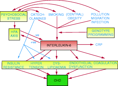
Fig. 3.4
The relationship of obesity, stress, environmental factors and cardiovascular risk factors to interleukin-6 (Reproduced with permission of Elsevier from Yudkin et al. [12])
Multiple studies have demonstrated that elevated levels of CRP are a predictor of increased degrees of CAD, death from CAD, and stroke [14].
CRP Versus Highly-Sensitive CRP
Elevated levels of CRP are nonspecific and can occur in response to acute illness.
The degree of CRP elevation has been shown to correlate to risk of major adverse coronary events in patients suffering from an acute coronary syndrome and non-coronary infections and inflammatory diseases where levels of CRP can be greater than 10 mg/L.
However, there are significant inter-individual variations in CRP levels even within previously reported normal ranges among apparently healthy individuals who may be detected using a more sensitive assay.
For the prediction of cardiovascular events, the use of highly-sensitivity CRP (hsCRP) is the preferred assay for the measurement of CRP < 10 mg/L in patients who have no history of coronary or peripheral arterial atherosclerotic disease, independent of other coronary artery disease risk factors.
Risk categories associated with hsCRP [15]:
Normal: <1 mg/L
Intermediate risk: 1–3 mg/L
High risk: >3 mg/L
For patients with acute illness, values >10 mg/L can be expected. In these patients, levels should be reevaluated 2 weeks or more after the acute illness resolves [13].
Highly-Sensitive CRP for Primary Prevention
There have been numerous studies evaluating the potential for incremental improvement in cardiovascular risk prediction through the use of hsCRP testing among patients without known CHD:
In the EPIC-Norfolk prospective population study 1993–2003, British individuals with an elevated hsCRP, regardless of LDL levels and other cardiovascular risk factors, were at significantly increased risk for coronary and vascular events, and hsCRP was among the strongest predictors of CAD incidence and mortality (Table 3.3) [16].
Table 3.3
Fully adjusted risk estimates for incident coronary artery disease for various cardiovascular risk factors in EPIC-Norfolk
Cases/controls
All cases vs. controls 1,108/2,164
Fatal cases vs. controls 338/649
Sex
Matched
Matched
Age
Matched
Matched
Smoking
2.13 (l.65–2.75); <0.0001
2.46 (1.55–3.91); 0.0001
Diabetes
4.26(2.61–6.93; <0.0001
2.61 (1.24–5.49); 0.01
BMI
1.74 (1.36–2.23); <0.0001
1.27 (0.82–1.96); 0.3
Systolic BP
1.56 (1.22–1.99); <0.0001
1.96 (1.24–3.11); 0.001
LDL-c
1.68 (1.33–2.14); <0.0001
1.41 (0.89–2.24); 0.1
HDL-c
0.57 (0.44–0.73); <0.0001
0.80 (0.49–1.30); 0.3
CRP
1.66 (1.31–2.12); <0.0001
2.92 (1.83–4.67); <0.0001
In apparently healthy women without clinically significant hyperlipidemia, hsCRP has been proven to be a strong predictor of the risk of future cardiovascular events [17].
Regardless of baseline total cholesterol levels, hsCRP is an independent predictor of first MI amongst men (Fig. 3.5) [18].
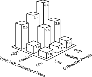
Fig. 3.5
Relative risk of first MI among apparently healthy men associated with high (>5.01), middle (3.78–5.01) and low (<3.78) tertiles of the total cholesterol-HDL ratio and high (>1.69 mg/dL), middle (0.72–1.69 mg/dL) and low <0.72 mg/dL) tertiles of CRP (Reproduced with permission from Ridker et al. [18])
hsCRP has been shown to improve risk prediction independently of standard cardiovascular risk factors, and hsCRP therefore may improve risk prediction compared to Framingham risk score alone.
Investigators have evaluated whether the incorporation of hsCRP, in addition to family history of premature coronary heart disease, may improve risk prediction as compared to the FRS:
The Reynolds Risk Score (RRS), a score that adds both hsCRP and family history of premature CHD to the Framingham risk score, was derivated and applied prospectively in both non-diabetic men and women without known CHD [19, 20].
These investigators showed that hsCRP and family history can modestly improve risk discrimination as compared to FRS alone and that this then leads to a significant percentage of patients being “re-classified” to either a higher or lower risk group (e.g., from intermediate risk by FRS to high risk).
Statins have been shown to reduce CRP levels regardless of LDL levels [21]:
The achieved level of CRP during statin therapy has been shown to be an additional predictor of benefit. In particular, achieved levels of CRP less than 1 mg/L identified a treated population with increased likelihood of atherosclerosis regression and greater reductions in event risk [22].
Recently, the JUPITER Trial [18] compared the use of the statin rosuvastatin (20 mg/day) to placebo amongst 17,802 statin-naïve patients (men >50 years-old; women >60 years-old) with LDL < 130, hsCRP > 2.0 and no history of diabetes, prior clinically apparent atherosclerotic cardiovascular disease (CVD), or significant chronic kidney disease:
This study demonstrated that treatment with rosuvastatin over a median of 1.9 years significantly reduced the risk of myocardial infarction, stroke, revascularization for unstable angina and cardiovascular death (combined endpoint adjusted hazard ratio 0.53; 95 % CI 0.40–0.69; p = 0.02), and overall mortality.
Based on the JUPITER Trial and the available literature to date, the 2010 ACCF/AHA Guideline for Assessment of Cardiovascular Risk in Asymptomatic Adults recommends measurement of CRP in select populations:
Class IIa: In men 50 years of age or older or women 60 years of age or older with LDL cholesterol less than 130 mg/dL; not on lipid-lowering, hormone replacement, or immunosuppressant therapy; without clinical CHD, diabetes, chronic kidney disease, severe inflammatory conditions, or contraindications to statins: Measurement of CRP can be useful in the selection of patients for statin therapy. (Level of Evidence: B)
Class IIb: In asymptomatic intermediate-risk men 50 years of age or younger or women 60 years of age or younger: Measurement of CRP may be reasonable for cardiovascular risk assessment. (Level of Evidence: B)
Class III:
In asymptomatic high-risk adults, measurement of CRP is not recommended for cardiovascular risk assessment. (Level of Evidence: B)
In low-risk men younger than 50 years of age or women 60 years of age or younger, measurement of CRP is not recommended for cardiovascular risk assessment [23]. (Level of Evidence: B)
CRP as a Predictor of CAD Severity and Secondary Prevention
In patients undergoing coronary artery angioplasty or surgical revascularization, baseline CRP has been found useful for predicting post-procedural adverse cardiac events.
Following elective or urgent percutaneous coronary intervention, patients with an elevated CRP (>3 mg/L) have been shown to have significantly higher incidence of myocardial infarction or death when compared to patients with a normal CRP level (6.1 % vs. 1.5 %, P < 0.0001). Repeat coronary revascularization was unrelated to CRP [24].
Among patients with unstable angina or non-ST segment elevation MI, 14-day mortality was predicted by elevated CRP (7.2 vs. 13 mg/L, P = 0.0038). Patients with an elevated CRP and troponin had the highest risk of mortality [25].
Following coronary revascularization for unstable angina, CRP has been shown to predict risk of coronary restenosis [26].
Limitations of hsCRP
The utility of hsCRP measurements is not without controversy and noted limitations. The normal hsCRP level is widely accepted; however, some disagree with such levels in the context of gender, ethnic, and genetic influences on CRP levels.
For patients with very low LDL, CRP level has not been related to the degree of atheroma progression by intravascular ultrasound [27].
There had been significant debate regarding the precise role that CRP played in the atherosclerotic process. Specifically, this debate centers on the question of whether CRP is a marker of risk or subclinical disease or is a causative risk factor involved mechanistically in atherosclerosis progression and clinical events (Fig. 3.6) [13, 28].
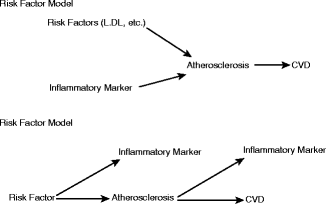
Fig. 3.6
Alternative models for the role of inflammatory markers in cardiovascular disease: risk factor or risk marker? (Reproduced with permission from Pearson et al. [13])
Despite data linking CRP to atherosclerotic plaque and vascular function [29], recent genetic studies strongly suggest that CRP is a risk marker rather than a direct risk factor. In a study evaluating large numbers of patients with various numerous single nucleotide polymorphisms in genes coding for CRP that result in significantly increased CRP levels, CRP does not appear to have a causative role in cardiovascular diseases [29]:
Among 10,276 patients, polymorphisms that resulted in markedly increased production of structurally normal CRP were not associated with an increase degree of ischemic vascular disease in this large cross-sectional analysis [30].
Oxidized LDL
Oxidized LDL (ox-LDL) is the product of lipid peroxidation from oxygen free radicals [33].
The modification of polyunsaturated fatty acids by oxidation with oxygen free radicals produces a reactive species that modifies both the lipid and protein components of LDL resulting in degradation of apo B-100 to peptide fragments that are further modified into oxidized fatty acids (Fig. 3.7) [34, 35].
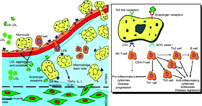
Fig. 3.7
Immune responses against oxidized LDL. LDL particles entrapped in the extracellular matrix of the artery wall become oxidized and activates an inflammatory response leading to infiltration of monocytes and T cells. The oxidized LDL is then taken up by macrophage scavenger receptors. While the cholesterol is stored in lipid droplets other structures in the oxidized LDL are processed for presentation on CD1 (lipids) and MHC class II molecules (peptides). In situations favoring maturation of naïve T cells into Th1 phenotype, this results in inflammation and disease progression. In contrast, if T cells are stimulated to mature into Th2 or regulatory T cells, inflammation and disease progression may not progress (Reproduced with permission of Elsevier from Steinbrecher et al. [34])
As the oxidized LDL and its byproducts accumulate on the arterial wall, they activate the inflammatory response that recruits T cells and monocytes. Monocytes develop into macrophages that ingest oxidized LDL, resulting in foam cells which initiate the cholesterol plaque as fatty streak [6, 35].
Chisolm et al. first reported in 1979 that ox-LDL was harmful to arterial walls and LDL related cytotoxicity was inhibited by HDL [36].
Oxidized LDL is atherogenic and has been shown to be potentially useful in the diagnosis, management, and prediction of stable and acute CAD.
Primary Prevention and Oxidized LDL
Circulating levels of oxidized LDL is a predictor of future CAD events. In a male population without a history of known CAD or diabetes and with adjusted traditional risk factors, ox-LDL was the strongest predictor of future CAD events compared to traditional risk factors and lipid profile (Fig. 3.8) [37].
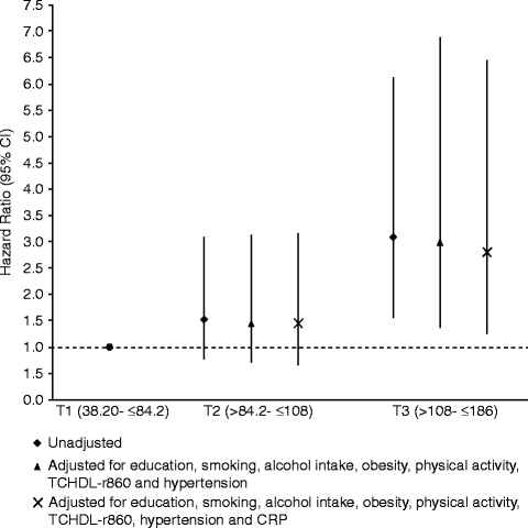
Fig. 3.8
Hazard ratios of coronary heart disease events according to tertiles (T1, T2, T3) of oxidized LDL(U/L) at baseline (Reproduced with permission from Meisinger et al. [37])
Elevated levels of ox-LDL independently predict increased prevalence and severity of subclinical atherosclerosis, as identified by ultrasound evaluation of the femoral and carotid arteries (Table 3.4) [38].
Table 3.4
IMT and plaque occurrence in the study group when divided in tertiles on basics of Ox-LDL
Tertiles of Ox-LDL
P for trend
Lowest (n = 129)
Middle (n = 128)
Highest (n = 129)
Ox-LDL U/L
61.5 ± 8.9
83.6 ± 5.8
113.3 ± 17.3
NT
IMT, mm
Common carotid artery
0.788 ± 0.120Related posts:
 Insights into the Natural History of Atherosclerosis Progression
 Positron Emission Tomography: Imaging Inflammatory Atherosclerosis
 Factors Contributing to the Development of Atherosclerosis
 Intravascular Ultrasound
 Noncontrast Cardiac CT for the Detection of Calcified Atherosclerosis
 Coronary Atherosclerosis Imaging by Coronary CT Angiography

Stay updated, free articles. Join our Telegram channel

Full access? Get Clinical Tree

 Get Clinical Tree app for offline access
