Nonunion of Proximal Humerus Fractures
Julie Y. Bishop
Evan L. Flatow
J. Y. Bishop: Mount Sinai School of Medicine, Department of Orthopaedic Surgery, New York, New York.
E. L. Flatow: Department of Orthopaedic Surgery, Mount Sinai Medical Center, New York, New York.
INTRODUCTION
Fractures of the proximal humerus are nondisplaced in the majority of cases and usually heal uneventfully with nonoperative treatment.15,27,29,41 Nonunions of the proximal humerus are uncommon and pose a very difficult problem to both the patient and the surgeon. For the patient, an established nonunion can be painful and functionally disabling. For the surgeon, treatment of these cases is extremely challenging, because various technical factors, such as soft bone, extensive scarring, and prior hardware, can limit the success of surgical treatment. However, successful treatment of a proximal humeral nonunion can be very rewarding when pain is abolished and functional use is restored to the patient.
PATHOETIOLOGY
Proximal humerus fractures are common, especially in elderly women.2,7,14,16,21,35 Nonunions, although rare, are more commonly seen after displaced two-part fractures or after cases in which open reduction and internal fixation is the initial treatment.6 Patients primarily present with a considerable amount of pain and varying degrees of functional loss. Patients are rarely able to perform their normal activities of daily living, and pain, which is often present at rest, is generally exacerbated by any attempt to use the extremity. The causes of nonunion can be divided into factors that are unique to the fracture site, factors associated with the first method of treatment, and systemic and medical factors.
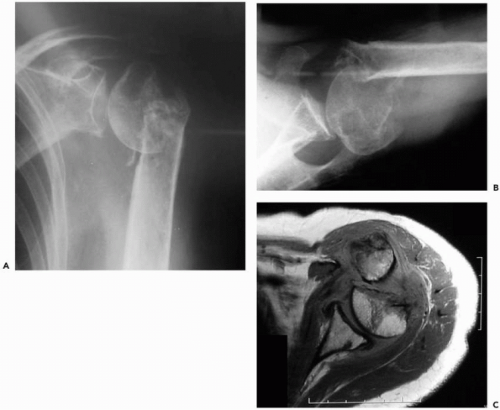
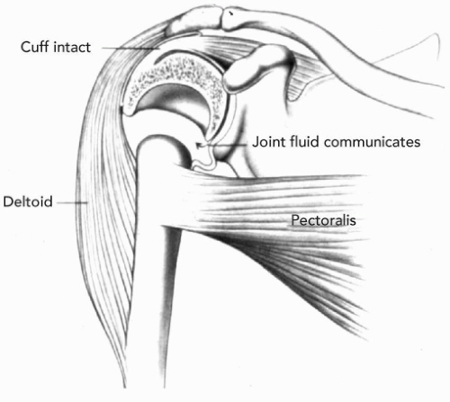
The weight of the arm causes a distraction force across the fracture site, which is compounded by the more cortical distal fragment that has poorer healing qualities compared to the more cancellous proximal fragment. A soft and cavitated head further decreases the healing potential of the bone in this area (Fig. 16-1). Soft-tissue interposition, such as the biceps tendon, can prevent fracture healing as well. Synovial fluid from the adjacent joint can communicate with the fracture site and limit the formation of hematoma and subsequent healing.
If the shaft is not properly reduced, the pectoralis major can continue to pull the shaft anteriorly, and the head can be further displaced through the pull of the rotator cuff. Improper immobilization can also create traction across the fracture site, also impeding bony apposition and healing. The arm must be immobilized across the front of the body, with the elbow anterior to the midline of the body in the coronal plane. This will neutralize the muscular forces across the fracture site (Fig. 16-2). Range-of-motion exercises, which are started before healing has begun, can also lead to nonunion of the fracture site. The upper extremity must move as a unit before any therapy is begun.
Finally, systemic disease and polytrauma are factors that may compromise a patient’s ability to heal a fracture. Elderly patients can have severe osteopenia, complicated by nutritional deficiency and/or metabolic bone diseases, again hindering the ability to heal fractures. These medical
issues should be recoginized and addressed prior to subsequent surgery and a referral made to an internist, if necessary. In the series by Healy et al., 16 of the 25 patients had significant medical illnesses, and the results of treatment in this group were unsatisfactory overall.14
issues should be recoginized and addressed prior to subsequent surgery and a referral made to an internist, if necessary. In the series by Healy et al., 16 of the 25 patients had significant medical illnesses, and the results of treatment in this group were unsatisfactory overall.14
CLINICAL EVALUATION
The amount of pain and functional losses that a patient has must be carefully assessed. Because of the potential for increased morbidity associated with attempted treatment of this problem, conservative management may be indicated for the minimally symptomatic patient. A complete review of all prior records, especially any operative reports, is essential.
On physical examination, false motion may occur through the nonunion site, and thus passive “shoulder” motion may seem excellent, whereas active motion is generally poor due to a flail arm. In fibrous nonunions, there may be some stability, and motion may be limited by shoulder stiffness. A careful neurovascular examination must be performed, especially if operative intervention is being considered. Neurologic damage may have occurred at the time of the original injury, or at the initial surgery, if one was performed. Special attention should be directed to the axillary nerve and the status of the deltoid.
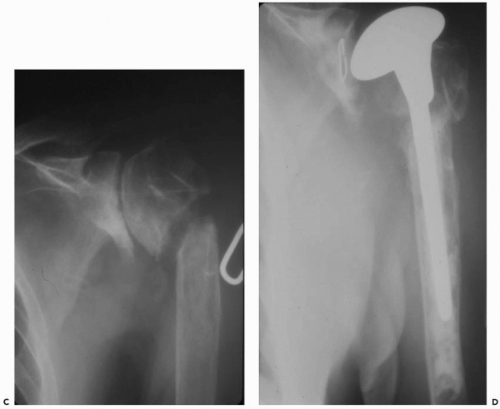
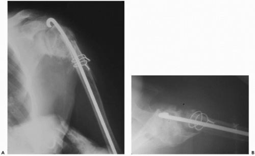 Figure 16-3 Infected nonunion after prior Ender’s rod fixation of the surgical neck fracture. (A) Anteroposterior (AP) and (B) axillary views. (continued) |
The status of the rotator cuff is often difficult to assess, because pain and weakness due to the original injury and subsequent nonunion may be significant. Overall disuse of the extremity may contribute to deltoid and periscapular muscular atrophy.
If the patient had prior surgical intervention, the possibility of sepsis must always be ruled out (Fig. 16-3). The skin should be carefully examined for any draining sinuses or other evidence of infection. When in doubt, appropriate lab work should be ordered and an aspiration performed with or without appropriate nuclear medicine imaging studies.
Finally, it is also very important to take into account the physiologic state of each patient and that patient’s ability to heal a fracture in light of his or her medical conditions.
RADIOGRAPHIC EVALUATION
Adequate radiographic evaluation is a necessity, and the standard views are always obtained: anteroposterior in the plane of the scapula, a scapular lateral or “Y” view, and an axillary view. This should be sufficient to judge the position of the fracture fragments, the extent of bone loss, and the quality of the bone present. Computed tomography (CT) scanning may be helpful, if radiographs are inconclusive. This is more common when there is malunion of the greater tuberosity and nonunion of the surgical neck in a three-part proximal humerus fracture. Magnetic resonance imaging (MRI) is useful to demonstrate any soft-tissue abnormalities associated with the biceps tendon, rotator cuff, deltoid, or labrum.
TUBEROSITY FRACTURE NONUNION
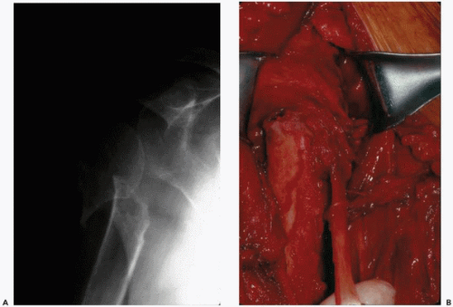
Tuberosity nonunions are rare unless markedly displaced; they usually heal to something and are more likely to develop a malunion. Gartsman and Taverna reported on a greater tuberosity nonunion, which was repaired arthroscopically, along with a concurrent rotator cuff tear.13 If necessary, greater and lesser tuberosity nonunions can be repaired with the same techniques utilized for acute fractures (Fig. 16-4). However, a long-standing nonunion of a tuberosity may be quite retracted and adhesed to the soft tissues, necessitating an extensive release for mobilization of the fragment. Surgical indications are similar to those for malunions and include significant displacement of the tuberosity and concurrent functional deficit caused by pain or significant limitation of range of motion.
SURGICAL NECK NONUNION
Surgical neck nonunions are more common than tuberosity nonunions and occur with or without tuberosity malunion, avascular necrosis of the humeral head, or posttraumatic glenohumeral arthritis41 (Fig. 16-5). Indeed, surgical neck nonunion is more likely, if there is glenohumeral stiffness, because motion is shifted to the fracture site. Operative treatment of a surgical neck nonunion is a challenging undertaking, because there can be extensive bone resorption,
including a portion of the head by the pseudoarthrosis. In addition, the remaining bone is often osteoporotic and holds fixation poorly. Neer25 noted in 1983 that there were only 29 reported cases in the literature, and then his paper added an additional 50 cases. Overall, the reported treatment results from the studies, subsequently in the literature, have been very mixed. Many types of treatment methods have been described6,14,18,24,31,33,36,37 that consist primarily of intramedullary fixation, open reduction and internal fixation, hemiarthroplasty, and “skillful neglect” or nonoperative treatment. Although some authors report reasonable results with humeral head replacement,1,4,9,30,38 others have noted more reliable functional results following open reduction and internal fixation (ORIF).3,4,8,30,31,33,36 In fact, one recent study recommends that replacement arthroplasty for surgical neck nonunions requiring greater tuberosity osteotomy be abandoned as a treatment option due to poor outcome.4 Each method will be reviewed, followed by the senior author’s treatment of choice.
including a portion of the head by the pseudoarthrosis. In addition, the remaining bone is often osteoporotic and holds fixation poorly. Neer25 noted in 1983 that there were only 29 reported cases in the literature, and then his paper added an additional 50 cases. Overall, the reported treatment results from the studies, subsequently in the literature, have been very mixed. Many types of treatment methods have been described6,14,18,24,31,33,36,37 that consist primarily of intramedullary fixation, open reduction and internal fixation, hemiarthroplasty, and “skillful neglect” or nonoperative treatment. Although some authors report reasonable results with humeral head replacement,1,4,9,30,38 others have noted more reliable functional results following open reduction and internal fixation (ORIF).3,4,8,30,31,33,36 In fact, one recent study recommends that replacement arthroplasty for surgical neck nonunions requiring greater tuberosity osteotomy be abandoned as a treatment option due to poor outcome.4 Each method will be reviewed, followed by the senior author’s treatment of choice.
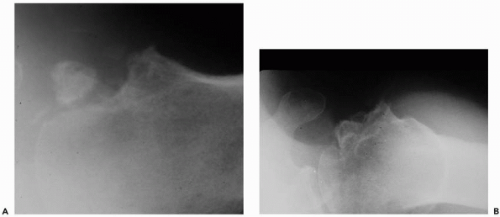 Figure 16-4 (A) Nonunion of a lesser tuberosity fracture, which was treated nonoperatively. (B) Axillary view after open reduction and suture fixation of the lesser tuberosity fragment. |
NONOPERATIVE TREATMENT
Stay updated, free articles. Join our Telegram channel

Full access? Get Clinical Tree


