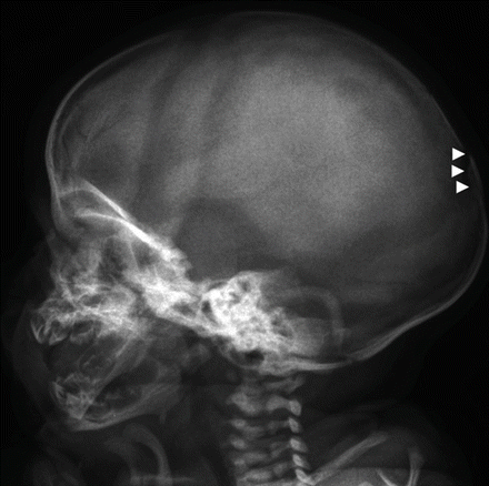
Skull
Anterior posterior (AP), lateral, and Towne’s view (the latter if clinically indicated)
Skull radiographs should be taken with the skeletal survey even if a CT scan has been or will be performed
Chest
AP including the clavicles
Oblique views of both of the sides of the chest to show ribs (‘left and right oblique’)
Abdomen
AP of abdomen including the pelvis and hips
Spine
Lateral: this may require separate exposures of the cervical, thoracic and thorocolumbar regions
If the whole spine is not seen in the AP projection on the chest and abdominal radiographs, additional views will be required
AP views of the cervical spine are rarely diagnostic at this age and should only be performed at the discretion of the radiologist
Limbs
AP of both upper arms
AP of both forearms
AP of both femurs
AP of both lower legs
Posteranterior view of hands
Dorsoplantar view of feet
Occasionally radio-isotope bone scanning may help when plain radiographs are equivocal for skeletal injury. They are particularly useful in rib, spinal and diaphyseal fractures, but not as useful for metaphyseal or skull fractures. Biochemical, haematological and genetic investigations should be considered to exclude NAI. These include bone profiles, markers of metabolic disease and parathyroid levels.
12.3 Specific Injuries (Table 12.2)
Table 12.2
Specificity of fracture types for paediatric non-accidental injury
Fractures with high specificity |
Metaphyseal fractures |
Rib fractures |
Scapular fractures |
Outer-end clavicle fractures |
Fractures of different ages |
Vertebral fractures or subluxation |
Digital injuries in non-mobile children |
Bilateral fractures |
Complex skull fractures |
Frequent fractures but with low specificity |
Mid-clavicular fractures |
Simple linear skull fractures |
Single long-bone fractures |
12.3.1 Skull Fractures
Accidental skull fractures in young children are uncommon. It is unlikely that a young child will sustain a skull fracture in a fall on the head of less than 1 m. In NAI, skull fractures tend to involve more than one bone, and hence to cross suture lines. Suspicion should also be raised in depressed fractures and so-called “growing” fractures, when the fracture gap increases with time (Fig. 12.1).


