Site
Age in years
Capitellum
1
Radial head
3
Medial epicondyle
5
Trochlea
7
Olecranon
9
Lateral epicondyle
11
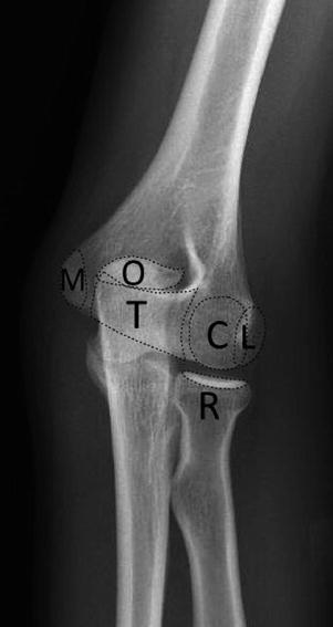
Fig. 14.1
Left elbow AP oblique with external rotation in a 15-year-old demonstrating the fused elbow ossification centers. C capitellum, R radial head, M medial epicondyle, T trochlea, O olecranon, L lateral epicondyle
The radiocapitellar relationship is important to note when assessing the elbow. On the lateral view, the anterior humeral line and the radiocapitellar line are used to assess the elbow joint alignment. The anterior humeral line is a line drawn along the anterior cortex of the distal humeral metaphysis which should pass through the middle third of the capitellum (Fig. 14.2). If the anterior humeral line passes in the anterior third of the capitellum or anterior to the capitellum, then the capitellum is too posterior, indicating a distal humeral fracture. The radiocapitellar line is a line drawn through the radial neck which should pass through the capitellum (Fig. 14.3). If the radiocapitellar line does not pass through the capitellum, then there is a radiocapitellar dislocation.
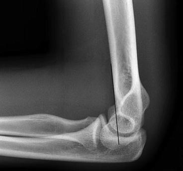
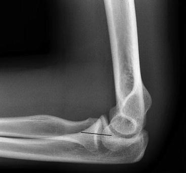

Fig. 14.2
Lateral view of the elbow with a normal anterior humeral line. The anterior humeral line is a line drawn along the anterior cortex of the distal humeral metaphysis which should pass through the middle third of the capitellum

Fig. 14.3
Lateral view of the elbow with a normal radiocapitellar line. The radiocapitellar line is a line drawn through the radial neck which should pass through the capitellum
Radius and Ulna
The primary radiographs of the radius and ulna are the AP and lateral views. For sufficient evaluation, the elbow and wrist should also be visible because there are important fractures that include dislocation components. A Monteggia fracture-dislocation is a fracture of the ulna shaft with an associated dislocation of the radial head. A Galeazzi fracture-dislocation is a fracture of the radius with a dislocation of the distal radioulnar joint.
Another common fracture is a Colles’ fracture. A Colles’ fracture is a fracture of the distal metaphysis of the radius associated with dorsal angulation and displacement of the distal radius typically seen in the older population or women with osteoporosis. In addition to inclusion of the elbow and wrist joints, care must be taken to avoid pronation or supination between views as either of these changes in position will change the orientation of the osseous structures and potentially obscure pathology.
Wrist
Radiographs of the wrist should include the carpal bones as well as the metacarpals, distal radius, and distal ulna. AP, lateral, and oblique views of the wrist are the standard views obtained. In the setting of suspected fracture of a specific bone, particularly the scaphoid, dedicated views of the specific bone can be requested to improve evaluation.
The carpal alignment can be assessed by evaluating the three carpal arcs on the AP view. If there is disruption of the carpal arcs, then a fracture or ligamentous injury is suggested. The first arc traces the proximal convexities of the scaphoid, lunate, and triquetrum. The second arc traces the distal concave surface of the scaphoid, lunate, and triquetrum. The third arc traces the main proximal curvatures of the capitate and hamate. See Fig. 14.4 which demonstrates the normal carpal arcs. In addition to the carpal arcs, specific ligamentous injuries can be suggested on the AP view. In the setting of a scapholunate ligament injury, then there can be widening of the scapholunate joint. In the setting of a fracture of the ulnar styloid, then there may be injury to the triangular fibrocartilage (TFCC). If a ligamentous injury is suspected, MRI should be considered.
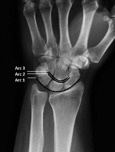

Fig. 14.4
AP view of the wrist with normal carpal alignment demonstrating the three carpal arcs. Arc 1 traces the proximal convexities of the scaphoid, lunate, and triquetrum. Arc 2 traces the distal concave surfaces of the scaphoid, lunate, and triquetrum. Arc 3 traces the main proximal curvatures of the capitate and hamate
The most common wrist dislocations are the perilunate and lunate dislocations which can be assessed on the lateral view. Figure 14.5 demonstrates the normal lateral wrist alignment. If the capitate is centered over the radius with the lunate tilted towards the palmar surface, then there is a lunate dislocation. If the lunate is centered over the distal radius with the capitate located dorsally, then there is a perilunate dislocation.
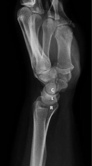

Fig. 14.5
Lateral view of the wrist with normal alignment of the distal radius (R), lunate (L), and capitate (C)
Hand
To evaluate the hand, AP, lateral, and oblique radiographs are typically obtained. If individual phalangeal injuries are suspected, then dedicated radiographs of the designated phalanx should be ordered. The phalanges can be designated by name or number. Using the numbering system, the thumb is designated the first digit, the index finger is designated the second digit, and so on.
Pelvis and Hip
In the setting of a patient with a history of trauma and pain during weight bearing, images of both the pelvis and hip should be considered. Radiographs of the pelvis and hip starts with an AP view of the pelvis. After the pelvis has been imaged, AP and frog leg lateral views of the affected hip should be obtained. Weight-bearing AP views of the hip or pelvis can be subsequently considered if the initial images are negative. Keep in mind that hip pain can be referred from the lumbar spine or from extra-articular causes.
Femur
Upper leg complaints may be due to pathology of the hip, femur, knee, or lumbar spine. When the appropriate history and physical examination indicates the femur as the pathological source, AP and lateral views of the femur can be added to images of the hip or less commonly images of the knee.
Knee
Radiographs of the knee typically include an AP, lateral, and sunrise view. The initial views are typically obtained supine, but weight-bearing AP and PA can be subsequently obtained to further evaluate the medial and lateral compartments. The AP view primarily is used to evaluate the medial and lateral femoral compartments. The lateral view helps determine if a joint effusion is present. The sunrise view is used to primarily evaluate the patellofemoral compartment.
Stay updated, free articles. Join our Telegram channel

Full access? Get Clinical Tree


