Musculoskeletal infection

6 Musculoskeletal infection


Cases relevant to this chapter


12, 23, 26, 27, 32–33, 68, 71, 75, 79, 91


Introduction


Various musculoskeletal tissues can become infected, giving rise to a variety of clinical problems (Table 6.1). The underlying causes and the basic principles of management are similar, irrespective of the tissue concerned. Here we will look at why and how tissues become infected, consider the clinical presentation, and describe the principles governing management of these conditions. Specific types of infection will then be considered in greater detail.


Pathology of musculoskeletal infection


In this section we are concerned exclusively with bacterial infection. In some parts of the world, fungal infections are of importance, but, even so, these are relatively uncommon.


Musculoskeletal infections occur either by haematogenous spread (that is, via the bloodstream) or by direct inoculation through open wounds or skin lesions. Bacteria may come from an established infection elsewhere (for example, in the respiratory tract) or may simply be circulating in the bloodstream.


The presence of bacteria in the circulation (bacteraemia) does not usually lead to established infection either in the bloodstream (septicaemia) or in a local site, such as a joint. It is not uncommon for bacteria to circulate in the blood without being able to establish a focus of infection. Infection occurs when the infecting organism is present in sufficient numbers and with sufficient virulence to overcome the body’s defence mechanisms. These latter include physical barriers, the non-specific inflammatory response as well as specific immunological responses. There is, thus, a balance between host resistance and infection. Organisms that are normally considered to be of low virulence can cause infections when the host’s immune protection is compromised.


Certain tissues and regions are more susceptible to bacterial colonization than others. These include the metaphyseal regions of the long bones. This is attributed to the vascular arrangement in these areas, where sharp loops of capillaries predispose to bacterial deposition. Other susceptible tissues include any bone or muscle that has been damaged or otherwise has an impaired blood supply.


The commonest organism involved in musculoskeletal infection is Staphylococcus aureus, followed in frequency by species of Streptococcus. Haemophilus influenzae infections used to be common in children prior to the introduction of immunization against this organism. Other organisms are associated with the specific infections described below.


Clinical presentation of musculoskeletal infection


In general terms, acute infection is accompanied by a systemic response, including fever, malaise and tachycardia, although these features may not be evident in the neonate. With increasing severity of infection and the presence of bacterial products in the bloodstream, there may be rigors and signs of shock (septic shock). As an abscess develops, the fever is classically a swinging fever, with peaks of high temperature corresponding to the release of bacterial toxins in the bloodstream. The site of the infection will show the classical signs of inflammation, with local warmth, redness, swelling and tenderness. These signs will be most obvious in infections of tissues near the surface and, conversely, will be difficult to elicit in deep sites, such as the hip joint. The patient will complain of pain and will be reluctant to move the affected part (so-called pseudo-paralysis). Pseudo-paralysis in the infant is suspected when there is lack of spontaneous movements of a limb; this is often the only complaint the mother may report.


Typically, the white cell count will be raised, with a neutrophilia in acute bacterial infection. Inflammatory markers, such as the erythrocyte sedimentation rate (ESR) and the acute-phase protein, C-reactive protein (CRP), will be raised. These markers may be useful in monitoring the progress of the treated infection.


The indications for diagnostic imaging vary according to the specific types of musculoskeletal infection and will be described below.


Principles of management of musculoskeletal infection


As far as possible the causative organism should be identified and its antibiotic sensitivities confirmed. Blood cultures may be positive during a bacteraemic phase, but ideally material from the infection site should be obtained for culture and sensitivity before any antibiotic therapy is started. However, it must be emphasized that in a proportion of patients no organisms may be cultured and in such situations treatment should not be withheld.


In the early stages of infection, antibiotics alone may be sufficient to control and eradicate the infection. Initially, these should be administered intravenously to obtain adequate levels of antibiotic in the tissues concerned.


The affected part should be rested and splinted if appropriate, and analgesia prescribed as necessary. Antibiotics alone will not cure musculoskeletal infection at any site once abscess formation has become established, or where pus is accumulating in significant volumes. Under these circumstances drainage is indicated. Simple aspiration and washout of an involved joint may be all that is necessary, but open surgery and more radical measures will be required for bone infection.


A serious musculoskeletal infection in a child may be the first manifestation of diabetes mellitus, so blood glucose levels should be checked.


In infants careful examination of other joints and systems may identify other concomitant sites of infection.


Following an appropriate period of rest and protection, mobilization, with the help of physiotherapy, will be necessary to restore range of joint movement and muscle power, and minimize osteoporosis in affected bones.


Specific infections


Acute septic arthritis


Infection of a joint may occur by blood-borne infection, by direct penetrating injury (this may take the form of a surgical procedure on the joint) or by spread of infection from an osteomyelitic focus in an adjacent bone. Over all age groups the commonest organism is S. aureus. In sexually active adults, the commonest cause is Neisseria gonorrhoeae, the causative organism of gonorrhoea. About half of all patients with septic arthritis are under the age of 3 years. Across all age groups, the commonest joint affected is the knee, but in infants and young children septic arthritis of the hip is more common.


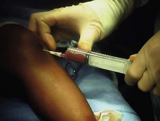
The clinical presentation is of pain and loss of movement in a joint together with fever and malaise. Prompt diagnosis and treatment is necessary, as articular cartilage does not survive long in the presence of a tense effusion of pus. A high index of suspicion for infection is required in all cases of an acutely swollen and painful joint, particularly in a young child. Aspiration of the joint (Fig. 6.1) may not yield the causative organism, and diagnostic imaging modalities such as radiography and ultrasonography will simply confirm the presence of a joint effusion. However, the clinical picture together with abnormally high values for the inflammatory markers should be sufficient to make the provisional diagnosis of infection. After blood cultures have been obtained, ‘best guess’ intravenous antibiotic treatment must be started and the joint drained and washed out with copious quantity of saline. At the hip, this may require open surgery, especially if the infection has become established and septation has occurred (subdivision of the abscess cavity by fibrin bands).


< div class='tao-gold-member'>

Stay updated, free articles. Join our Telegram channel

Jul 12, 2016 | Posted by in RHEUMATOLOGY | Comments Off on Musculoskeletal infection

Full access? Get Clinical Tree

Get Clinical Tree app for offline access