Fig. 26.1
Sixty-eight year-old female with history of endometrial carcinoma. Lateral radiograph of the right foot demonstrating and aggressive appearing lytic lesion involving the body of the calcaneus

Fig. 26.2
AP radiograph of the right foot

Fig. 26.3
Coronal CT image of the hindfoot demonstrating a lytic calcaneal lesion with disruption of the medial wall

Fig. 26.4
Sagittal CT image of the hindfoot illustrating the anterior to posterior extent of the lesion
Anatomic Locations
Hindfoot
Surgical management is directed by several factors including size of the lesion, bone(s) affected, location within the bone, and health of the soft tissue envelope. The hindfoot can be safely approached from either the lateral or medial side depending on tumor location. Experience from the treatment of calcaneus fractures has highlighted the need for meticulous handling of the soft tissue envelope when utilizing a lateral-based approach. This is particularly true when external beam radiation has been used as an adjunct to local tumor control. In patients with poor tissue perfusion and limited potential for wound healing (vascular disease, diabetes, malnutrition, fluid imbalance), strong consideration for non-operative management should be given.
Intra-lesional resection or curettage is undertaken with margin expansion utilizing power burr and/or adjuvants, such as argon beam or liquid nitrogen, as deemed necessary. Excessive resection of adjacent cortical or cancellous bone is not indicated and can have a negative impact on subsequent reconstruction. Following tumor resection, bony reconstruction is generally achieved with bone cement augmented with screw or wire fixation when possible (Figs. 26.5 and 26.6). Non-contained defects (those that involve loss of the normal cortical boundaries) may be supplemented with low profile, specialized plating systems to prevent cement extrusion and re-establish normal cortical boundaries. These techniques generally allow for early weight bearing activities while avoiding complications inherent with bone graft use such as nonunion or infection. Because of the abnormal biologic environment and limited healing potential, large structural bone grafts or bone graft fillers are not generally recommended.

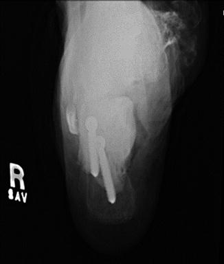
Fig. 26.5
Post-operative lateral radiograph of the hindfoot showing cement and screw reconstruction following intra-lesional resection of the metastatic lesion
Fig. 26.6
Post-operative Harris view of the calcaneus
Midfoot
Metastatic lesions involving the tarsal bones or proximal metatarsals are treated similarly to lesions found in the hindfoot. Due to size and anatomic constraints, use of screw or pin augmentation within individual bones is limited. It may be necessary, however, to consider transtarsal or metatarsal fixation in cases of large, non-contained lesions with significant bone loss. Dorsal, medial, or lateral approaches can be used safely depending on the particular site of involvement. Again, meticulous handling of the soft tissue envelope is paramount to avoid wound healing complications, dehiscence, and infection.
Forefoot
Lesions involving the metatarsals and phalanges are less commonly seen and often require no surgical intervention. When surgery is deemed necessary (often due to nonunion of a pathologic fracture and/or recalcitrant pain), consideration for toe or ray amputation should be given. These procedures are generally well tolerated with little effect on patient function and reliably relieve patient pain and discomfort. A notable exception would be amputations involving the first ray or great toe in which balance, walking, and foot alignment may be adversely affected.
Related posts:


Stay updated, free articles. Join our Telegram channel

Full access? Get Clinical Tree


