Fig. 3.1
Normal meniscus
The menisci enhance the stability of the knee joint by deepening and hence increasing the contact area between the femoral condyles and the tibial plateau. They help dissipate the contact hoop forces evenly across the articular surface. By virtue of the nerve endings in their anterior and posterior thirds, they contribute to the proprioception in the knee joint. The menisci also aid in lubrication in the knee joint [1, 2].
3.1.1 Embryology
In the developing embryo in the 28th–32nd day stage, the lower limb buds form opposite the lower lumbar and upper sacral segments. Gardner and O’ Rahilly [3], Mc Dermot [4] and others provided detailed descriptions of the prenatal development of the knee joint. However, they largely described the embryonic development of the joint (i.e., prior to three gestational months) [5]. Thus, the intrauterine development is divided into four stages:
1.
Formation of the uniform interzone:
The osteogenesis of the long bones starts from the 6th week onwards from primary ossification centres in the middle of the cartilaginous anlage of the long bones.
2.
Formation of the three-layered interzone:
At the end of the embryonic period (8 weeks, stage 23), the cells of the menisci are round and randomly arranged. The superficial cells begin to orient themselves parallel to the joint surface.
3.
Meniscal cell differentiation:
A layer of decreased cell density separates the menisci from the tibial plateaux. By 10 weeks, the densely celled menisci can be easily distinguished from a loose celled tissue peripherally which contains blood vessels.
4.
Collagenous matrix formation inside the menisci:
At 12 weeks, some blood vessels penetrate the peripheral third of both menisci. The orientation of collagen fibres becomes obvious at 14 weeks: it is parallel to the joint surface on the inner part of the menisci, and by 40 weeks, the vascularity of the entire meniscus is defined.
3.1.2 Discoid Meniscus
A congenital variant of the normal morphology of the meniscus is the discoid meniscus (Figs. 3.2 and 3.3). Smillie [6] suggested that this variation in structure is due to a failure of the foetal discoid form of meniscus to involute. He found in his series an incidence of around 6 %. A comprehensive study by Nathan and Cole [7] found 2.5 % of the menisci to be discoid. They are more common on the lateral side than the medial and rarely ever are found in both compartments. These can cause symptoms of snapping and popping in the knee in children, usually between the ages of 6 and 12 years.
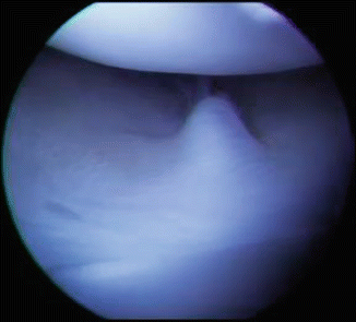
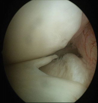

Fig. 3.2
Discoid lateral meniscus complete type

Fig. 3.3
Discoid lateral meniscus incomplete type
The entity of discoid lateral meniscus was first reported by Young [8] in 1889. In a small percentage of these patients, there is no attachment of the posterior horn to the tibial plateau and instead a continuous Wrisberg ligament. This absent insertion may have implications for meniscal stability, although instability of clinical significance is uncommon.
Multiple classifications have been proposed, the most commonly used being the Watanabe et al. [9] system of 1978. They described three different types:
1.
Complete, disk-shaped meniscus with a thin centre covering the tibial plateau
2.
Incomplete, semilunar-shaped meniscus with partial tibial plateau coverage
3.
Wrisberg-type, hypermobile meniscus resulting from deficient posterior tibial attachments
In 1998, Monllau et al. [10] identified a fourth type: the ring-shaped meniscus. Good et al. [11] proposed an interesting classification based on the discoid meniscal instability as anterior or posterior. In the vast majority of discoid menisci, the meniscus is simply an incidental finding on either MRI or arthroscopy and is asymptomatic.
3.1.3 Composition and Histology
Clarke and Ogden [12] are credited with the description of the postnatal development study of the human menisci, correlating anatomy and histology. The normal human meniscal tissue is composed of 72 % water, 22 % collagen, 0.8 % glycosaminoglycans and 0.12 % DNA [13]. Histologically, the menisci are fibrocartilaginous and are primarily composed of an interlacing network of collagen fibres interposed with cells, with an extracellular matrix of proteoglycans and glycoproteins. Type I collagen accounts for over 90 % of the meniscal collagen, the remainder consists of types II, II and IV [14]. Bullough et al. [15] found that the principal orientation of the collagen fibre is circumferential, to withstand tension. They also found some radially oriented collagen fibres which were believed to act as “ties” holding the circumferential fibres together.
This arrangement provides good tensile strength and aids in evenly distributing forces across the articular surface [16]. The circumferential fibres absorb the compressive forces, whereas the radial fibres help prevent longitudinal splitting [17]. Disruption of the meniscal integrity leads to uneven loading of the joint cartilage, leading to early osteoarthritis [18]. Each meniscus is divided into three segments: anterior third, middle third (body) and posterior third. It is the outer 20–30 % which is vascular (red zone) and is supplied by the medial and lateral geniculate arteries, respectively. The inner two thirds of the menisci are relatively avascular (white zone). This distribution of blood supply determines the treatment of meniscal tears [19].
3.2 Role and Functions
Over the years, understanding the functions and roles of the menisci has changed dramatically, and since King’s paper in 1936 [20], numerous studies have shown that the menisci play important roles. They have now been recognised as key primary stabilisers and weight transmitters in the knee and primarily distribute the contact forces across the tibiofemoral articulation, which is achieved through a combination of the material, geometry and attachments of the menisci. Fukubayashi and Kurosawa [21] examined the intra-articular contact areas using a casting method employing silicone rubber and found that the menisci combined occupied 70 % of the total contact area within the joint. Walker and Erkman [22] also used casting technique in their study and deduced that under no load, contact occurred primarily on the menisci, but that with loads of 150 kg and more, the menisci covered between 59 and 71 % of the joint contact surface area.
The medial meniscus is the larger of the two and covers about 50 % of the medial tibial plateau. The anterior third is attached to the medial tibial spine just (7 mm) anterior to the anterior cruciate ligament (ACL) insertion. The posterior third attaches anterior to the attachment of the posterior cruciate ligament (PCL). Medially, it is attached to the femoral condyle and tibial plateau by means of the coronary ligaments which form the deep portion of the medial collateral ligament [23]. The peripheral capsular attachment of the medial meniscus is continuous. This relative immobility of the medial meniscus renders it at a higher risk of injury when compared to the lateral meniscus [24, 25].
The lateral meniscus, which is the smaller of the two menisci, covers about 70 % of the lateral tibial plateau. The anterior third attaches anterior to the tibial spine sharing some fibres with the ACL. The posterior third is attached just posterior to the tibial spine [23]. The peripheral capsular attachment is interrupted by the popliteus hiatus through which passes the popliteus tendon. The meniscofemoral ligaments attach the posterior third of the lateral meniscus to the lateral margin of the posterior medial femoral condyle. The anterior part is called the ligament of Humphrey, and the posterior one is the ligament of Wrisberg. It is more mobile than the medial meniscus and may displace up to 1 cm, which may explain why meniscal injuries occur less frequently on the lateral side [24, 25].
The discoid meniscus (Figs. 3.2 and 3.3) is a developmental anomaly in which the meniscus is thickened and is disk shaped. Discoid medial menisci are very rare with a reported incidence of 0.06–0.3 % [26, 27] of the general population. Discoid lateral menisci are more common with a reported incidence of 1.4–15.5 % [28]. The Koreans and Japanese have a higher incidence (16 %) than the Caucasians (5 %) [29]. They are of three types: partial, complete and the Wrisberg type [30]. Partial and complete variants are determined by the amount of coverage of the tibial plateau. They have normal tibial attachments and are stable. The Wrisberg type has no posterior capsular and tibial attachment. The only attachment is the meniscofemoral ligament of Wrisberg. These are highly mobile and unstable but fortunately relatively uncommon.
3.3 Meniscal Tears
The incidence of meniscal tears is about 61/100,000 persons per year in the United States [31] and 60–70/100,000 persons in Europe [32]. This is a highly estimated figure as many tears are asymptomatic and a similarly high number is seen in degenerative knees. Meniscal tears are more common in males (male: female = 2.5:1 to 4:1). Anterior cruciate ligament injuries are associated with almost a third of meniscal tears, more commonly the lateral meniscus in the acute injury setting [33, 34].
Meniscal tears can occur as a result of acute events and also from chronic degeneration. Meniscal tears usually occur secondary to axial and rotational loads, and activities in which sudden stopping and turning movements occur lead to a majority of tears. Instability increases the risk of meniscal tear, and a significant number of tears occur with anterior cruciate ligament injuries. 32 % of meniscal tears happen during a sports injury, 39 % during activities of daily living such as squatting and 29 % do not have any identifiable cause/event [33].
Meniscal injuries are usually accompanied by sudden onset of pain in the involved knee followed by a delayed onset of swelling, and tenderness is usually present in the involved compartment. Mechanical symptoms such as locking, catching and giving way are frequently seen. Joint effusion is present and pain is felt on deep flexion or on squatting. Special tests include joint line tenderness and the McMurray and Apley grinding test. Joint line tenderness is the most sensitive test with a sensitivity of 74 %. A positive McMurray test with a palpable clunk is very specific for a meniscal tear (98 % specificity) but has a sensitivity of only 15 %. A positive Apley grinding test has a specificity of 70 % and sensitivity of 60 % [35–37]. Symptoms of associated ligament injuries may also manifest. Chronic degenerative tears present with episodes of mechanical symptoms. On top of the underlying pain of the degenerative process in the knee, the patient may complain of locking and/or catching of the knee. An injury or twisting episode is not necessary to produce these symptoms. Magnetic resonance imaging (MRI) is the investigation of choice to detect meniscal tears. X-rays are often performed to rule out any osseous injury.
Meniscal tears are classified on the basis of tear location, tear pattern or by blood supply. On the basis of tear location, they are anterior horn tears, tears of the body, posterior horn tears and root avulsions. Tears classified on the basis of pattern of tear are flap tears (Fig. 3.4), radial tears, complex/degenerative tears (Fig. 3.5), longitudinal tears, bucket handle tears (Figs. 3.6, 3.7 and 3.8), parrot beak tears (Fig. 3.9) and horizontal tears [38]. Cooper et al. described a circumferential zone classification based on the blood supply of the meniscus. Zone 0 is the menisco-synovial junction, zone 1 is the outer third of the meniscus, zone 2 includes the middle third and zone 3 is the central third of the meniscus [39].
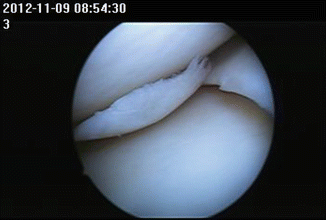
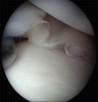
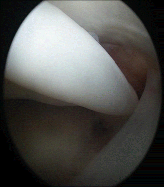
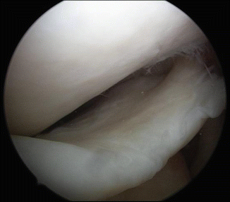
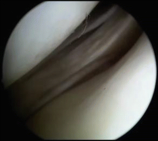
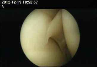

Fig. 3.4
Flap tear of medial meniscus

Fig. 3.5
Complex tear medial meniscus

Fig. 3.6
Bucket handle displaced in notch

Fig. 3.7
Displaced bucket handle tear of medial meniscus

Fig. 3.8
Undisplaced bucket handle tear of medial meniscus

Fig. 3.9
Parrot beak tear of medial meniscus
The International Society of Arthroscopy, Knee Surgery and Orthopaedic Sports Medicine (ISAKOS) formed a Meniscal Documentation Subcommittee in 2006 with the objective of developing a reliable, international meniscal evaluation and documentation system to facilitate outcomes assessment. In an interobserver reliability study, Anderson et al. [40] concluded that the ISAKOS classification of meniscal tears provided sufficient interobserver reliability for pooling of data from international clinical trials designed to evaluate the outcomes of treatment for meniscal tears.
Meniscal root tears (Fig. 3.10) differ distinctly from tears of the anterior or posterior horn of the meniscus. Meniscal roots are ligamentous attachments that help anchor the anterior and posterior horns of the meniscus to the tibial plateau. Root tears may or may not be associated with tears of the meniscus, and isolated root tears can occur with no meniscus tear itself. The meniscus is no longer anchored at the root attachment, leading to secondary extrusion. In this scenario, the meniscus will no longer perform its normal function of buffering the mechanical load imposed on the tibiofemoral joint, leading to tibiofemoral cartilage loss. In 2013 in a multicentre study, Guermazi et al. [41] concluded that isolated medial posterior meniscal root tear is associated with progressive medial tibiofemoral cartilage loss.
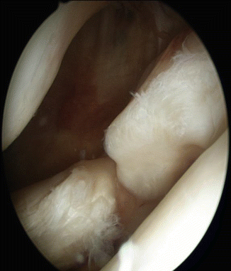

Fig. 3.10
Lateral meniscus root tear
Based on tear morphology, LaPrade et al. [42] classified meniscal root tear patterns into the following:
Type 1: Partial stable root tears
Type 2: Complete radial tears within 9 mm of the bony root attachment (further subclassified into types 2A, 2B and 2C, located 0 to <3 mm, 3 to <6 mm and 6 to 9 mm from the root attachment, respectively)
Type 3: Bucket-handle tears with a complete root detachment
Type 4: Complex oblique tears with complete root detachments extending into the root attachment
Type 5: Bony avulsion fractures of the root attachments
Takahashi et al. in their study aimed to identify factors on routine pulse sequence MRI associated with cartilage degeneration observed on T1ρ relaxation mapping. They concluded that posterior root/horn radial tears in the medial meniscus are particularly important MRI findings associated with cartilage degeneration observed on T1ρ relaxation mapping. Morphological factors of the medial meniscus on MRI provide findings useful for screening early-stage osteoarthritis.
The importance of anatomical repair of meniscal root tears was emphasised by LaPrade et al. [43] in another study. They concluded that the nonanatomic repair did not restore the contact area or mean contact pressures to that of the intact knee or anatomic repair. However, an anatomic repair produced near-intact contact area and resulted in relatively minimal increases in mean and peak contact pressures compared with the intact knee. Papalia et al. [44] stated that biomechanical and clinical studies demonstrate that surgical repair of acute, traumatic meniscal root injuries fully restores the biomechanical features of the menisci, leading to pain relief and functional improvement.
3.3.1 Meniscectomy and Osteoarthritis
Fairbank [45] as early as 1948 reported radiographic changes in the knee after total meniscectomy, describing osteophyte formation, femoral condyle flattening and narrowing of joint space. Baratz et al. [46] demonstrated a decrease in tibiofemoral contact area by 10 % and an increase in peak local contact stress by 65 % with partial meniscectomy, and these values increased to 75 % and 235 %, respectively, in situations where total meniscectomy was done. Tapper and Hoover [47] stressed the importance of joint instability as a predisposing factor for joint osteoarthritis instability following total meniscectomy. Hsieh and Walker [48], Levy et al. [49] and Hollis et al. [50] also stressed the important function of the menisci as secondary stabilisers, particularly the medial meniscus in providing resistance to A-P translation in ACL-deficient knees.
3.4 Treatment
Treatment of meniscal tears has evolved from conservative to surgical, from open to arthroscopic technique and from total meniscectomy to partial meniscectomy and meniscal repair. Meniscal allograft transplant has also evolved as a salvage procedure to replicate the meniscal function.
3.4.1 Non-operative Treatment
Non-operative treatment is appropriate for minimally displaced degenerative tears and consists of activity modification, physical therapy, local ice packs and anti-inflammatory medication. Once the acute pain subsides, the patients are prescribed range-of-motion (ROM) exercises, and after attaining full ROM, any muscle imbalance is corrected [51]. If symptoms fail to settle, then arthroscopic surgery can be considered but is usually unnecessary.
3.4.2 Meniscectomy
Historically, the surgical treatment of an injured meniscus was open complete excision. Bland Suttons in 1897 had described the menisci as functionless remnants of intra-articular leg muscles. McMurray [52] advocated a total meniscectomy for any meniscal tear and had gone to the extent of stating that “a far too common error is shown in the incomplete removal of the injured meniscus”. This philosophy was shown to be detrimental to the articular cartilage as described by Fairbanks in 1948, who described the changes in the knee joint following a meniscectomy which included ridge formation, narrowing of the joint space and flattening of the femoral condyle [53]. Further studies delineated the importance of preserving the meniscus as demonstrated by decreased contact areas and increased peak contact stress after a partial and/or total meniscectomy [54, 55]. Post meniscectomy, 74 % of knees had at least one Fairbank change and 39.4 % had degenerative arthritis as compared to 6 % of the contralateral normal knee [55]. Results of lateral meniscectomy are worse than that of medial meniscectomy, and resection of both menisci produced worse results than excision of a single meniscus. With the evolution of arthroscopic techniques, in the present day, almost all procedures related to the meniscus are done arthroscopically.
Meniscal tears in young patients and those associated with ACL tears should be repaired if at all possible. If the meniscus tear is complex and in an avascular zone, then it has no potential for healing, and a partial meniscectomy should be done. During a partial meniscectomy, unstable fragments should be removed to create a smooth transition, thus maintaining a functional meniscus. The short-term results of partial meniscectomy are excellent [56, 57]. Every attempt must be made to preserve as much of the meniscus as possible.
Stay updated, free articles. Join our Telegram channel

Full access? Get Clinical Tree


