Upper extremity injuries are more prevalent in obese people than in nonobese people after low-energy falls. Because splinting and casting are inefficient methods of stabilization in the setting of obesity, internal fixation provides stability for mobilization and realignment. Morbid obesity adversely affects positioning, surgical exposures, and complications associated with operative fixation. Avoiding short cuts and complications, morbidly obese patients should be able to return to normal functioning.
Health care costs are rising and represent 17.9% of the United States Gross Domestic Product in 2009. Representing almost 10% of all medical spending in 2008, obesity is a major component of health care costs. In the United States, obesity prevalence continues to increase over the past 40 years and is associated with decreased national productivity, medical expenditures, comorbidities, and perioperative complications. Obese individuals have up to 48% greater risk of injury, incurring higher rates of trauma, and lower extremity fractures in general. Specifically, obese people have an almost 2-fold increased chance of upper extremity injury as compared with those who are nonobese. Obese patients have a higher risk of sustaining a displaced elbow fracture than normal-weight patients.
This article discusses the treatment options and recommendations of upper extremity injuries in the setting of morbid obesity.
Shoulder injuries
Shoulder injuries can be subgrouped into clavicular, scapular, and proximal humeral fractures. The treatment of clavicular and scapular injuries in obese patients is similar to that in nonobese patients.
Problems arise with the combination of morbid obesity and unstable displaced proximal humeral fractures. Operative treatment options for displaced proximal humeral fractures are controversial. When combined with morbid obesity, fixation options can be problematic. A potential fixation option is closed reduction/percutaneous fixation. The 2.5-mm terminally threaded Schanz pins (Synthes, Paoli, PA, USA or Zimmer, Warsaw, IN, USA) are available in 250 or 300 mm length. However, swelling associated with fracture hematoma and obesity creates arm girth wider than commercially available pin length, restricting use of this procedure until swelling abates or abandoning the procedure.
Open reduction internal fixation via a deltopectoral interval or an extended lateral deltoid-splitting approach requires a large extensile incision. Because the proximal humeral plate is applied lateral to the biceps groove, the standard deltopectoral approach can limit unobstructed plate application secondary to the muscular interval being anterior to the plate location and to the depth of the dissection. The newly modified extended anterolateral acromial approach is probably the best suited for unobstructed plate application. In morbidly obese patients, the short drill sleeve and bit lengths can encumber minimally invasive plate application via submuscular plating.
Another option for salvage of displaced unstable proximal humeral fractures is arthroplasty. The problem with the deltopectoral approach and insertion of a proximal humeral arthroplasty stem is arm adduction and extension. Obesity, pendulous breasts, and wide girth all deter in line insertion of the arthroplasty stem within the humeral diaphysis. Having the patient placed onto the lateral aspect of the table with slight reverse Trendelenburg positioning can facilitate extension and adduction arm positioning. The surgeon must be prepared for all treatment methods concerning proximal humeral fixation.
Author’s Preferred Method
Since utilization of the upper extremity for weight bearing or getting out of a chair is common with morbid obesity, the author prefers locking plate fixation of proximal humeral fractures to insure stable fixation, avoid pin tract infections, and enhance mobilization. For unstable, dysvascular, and nonreconstructable fractures requiring arthroplasty, the author prefers a radiolucent table in reverse with the patient in a 30° reverse Trendelenburg and in a far ipsilateral lateralized position for positioning.
Humeral diaphyseal injuries
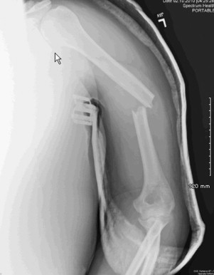
Most of the humeral fractures can be successfully treated with closed techniques. A typical treatment algorithm is initiated with humeral realignment and reduction maintained with a coaptation splint extending from under the axilla to above the shoulder. Once swelling and pain subside, the coaptation splint is changed to a fracture or functional brace. Functional bracing relies on hydrostatic forces to maintain fracture alignment and length. Bracing is maintained until stable callus formation develops and pain diminishes. Range of motion in and out of the brace is initiated based on fracture healing and stability.
Attempts at successful closed treatment of humeral diaphyseal fractures in obese patients are fraught with problems and complications. Splinting requires 3-point molding to achieve realignment ( Fig. 1 ). With excessive adiposity, bending moments are minimal, and alignment is unable to be achieved. Furthermore, in obese women more than in men, chest wall and breast size create a varus bending moment that is unable to be counteracted with traditional closed methods. Applying a rolled towel or bump on the medial aspect of the elbow outside of the splint will potentially offset the chest wall and breast forces. Once the initial pain and swelling subside, functional bracing begins. Functional bracing requires daily utilization that increases sweating, skin breakdown, and hygiene problems. Also, with the adiposity, hydrostatic forces are diminished if not absent. Therefore, closed methods of treating humeral diaphyseal fractures are insufficient.