Surgical Management of Stage 2 Adult Acquired Flatfoot
Jared M. Maker, DPM, AACFASa∗jmaker@coastalorthopedics.com and James M. Cottom, DPM, FACFASb, aFoot and Ankle Surgical Fellowship, Coastal Orthopedics and Sports Medicine, 6015 Pointe West Boulevard, Bradenton, FL 34209, USA; bCoastal Orthopedics and Sports Medicine, 6015 Pointe West Boulevard, Bradenton, FL 34209, USA
Adult acquired flatfoot deformity is a progressive disorder with multiple symptoms and degrees of deformity. Stage II adult acquired flatfoot can be divided into stage IIA and IIB based on severity of deformity. Surgical procedures should be chosen based on severity as well as location of the flatfoot deformity. Care must be taken not to overcorrect the flatfoot deformity so as to decrease the possibility of lateral column overload as well as stiffness.
Adult acquired flatfoot deformity
Posterior tibial tendon dysfunction
Flexor digitorum longus tendon transfer
Introduction
Adult acquired flatfoot deformity (AAFD) is a progressive as well as intricate disorder with multiple symptoms and numerous degrees of deformity.1,2 Johnson and Strom3 originally described 3 stages of AAFD that are practical when determining treatment options.1 Stage I patients complain of aching along the medial ankle without having secondary deformity. In stage II, pain increases in severity and distribution. The posterior tibial tendon (PTT) is elongated with secondary forefoot abduction as well as hindfoot valgus.3 Stage II has been further branched into stages IIa and IIb.4–6 Stage IIa has minimal abduction deformity through the midfoot with anterior-posterior (AP) radiographs revealing less than 30% talar head uncoverage. Patients with stage IIb reveal increased deformity on examination with AP radiographs revealing greater than 30% talar head uncoverage.4,5 Stage III has complete disruption of the PTT, with the patient having a fixed hindfoot valgus position along with secondary degenerative changes in the tritarsal complex.3 Myerson7 later described stage IV AAFD as valgus angulation of the talus along with degenerative changes within the ankle joint.
Patient physical examination
When performing a physical examination in the AAFD, the patient must be assessed in the relaxed stance position. When examining in the standing position, the forefoot may appear abducted in relationship to the rearfoot, causing the medial border to have a bulging and convex appearance with the lateral border shortened and concave.8 Forefoot abduction also is assessed from behind the patient with the “too many toes” sign. As an increase in severity of the deformity occurs, an increase in the number of toes laterally will be visualized.8,9 Commonly, a valgus deformity of the heel will be visualized as well.10
Following the stance examination, the reducibility of the deformity should be assessed. Single heel rise test is important to the examination when determining the function of the PTT with respect to stability of the arch. With a nonfunctioning PTT, the sagittal plane will reveal plantar flexion of the rearfoot on the forefoot. Minimal to no elevation of the heel will be visualized.10 When a patient is able to do a single heel rise, close examination of whether the heel shifts into varus must be assessed. The single heel rise test is positive if either the patient cannot elevate the heel off the ground or the heel does not shift into varus, staying in valgus during heel lift.4 Assessment of the reducibility of heel valgus can also be done with the double heel rise test.2
Range of motion (ROM) should be assessed at the ankle joint with the knee flexed and extended to determine if there is a gastrocnemius-soleus equinus or gastrocnemius equinus deformity. ROM should also be assessed at the subtalar joint.11 If the subtalar joint is unable to be reduced, reconstructive options may be inhibited.1,7,12 Assessment of PTT may reveal discomfort or fullness on palpation.11 Finally, PTT muscle strength of the affected limb should be assessed in relation to the contralateral side.10
Imaging
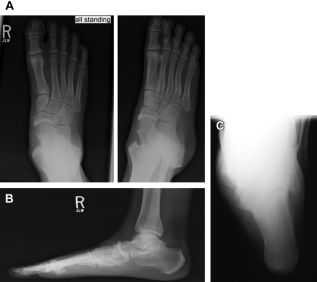
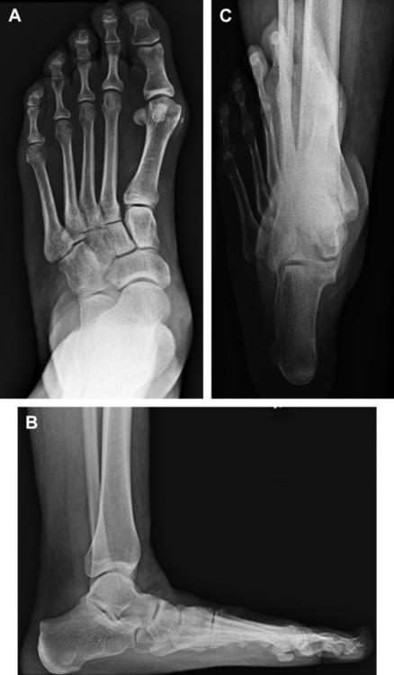
With stage II AAFD, radiographic analysis should be performed to assess the severity of the deformity. Typically, AP, lateral, medial oblique, ankle, and hindfoot alignment views are routinely done. Measurements on the AP view consist of talar first metatarsal angle, calcaneocuboid abduction angle, as well as talar head coverage when evaluating the amount of pronation and forefoot abduction. Measurements on the lateral view include talar first metatarsal angle and calcaneal inclination angle.2 The hindfoot alignment view allows the assessment of the frontal plane alignment of the calcaneus in relation to the tibia (Figs. 1 and 2).2,13,14


When evaluating the PTT, magnetic resonance imaging (MRI) will detect sheath inflammation, soft tissue resolution, and the current anatomic state of the tendon.15,16 Wacker and colleagues17 compared the muscle belly of the PTT and FDL tendon with MRI in patients with AAFD. Patients with complete rupture of the PTT revealed fatty infiltration within the muscle belly, therefore being nonfunctional. PTT without complete rupture showed 10.7% atrophy of the muscle belly and 17.2% hypertrophy of the FDL muscle belly. This study demonstrates the use of MRI with regards to surgical planning. With a complete rupture, augmenting the transferred FDL tendon to the PTT likely will not contribute much function.16
Conservative therapy
Conservative therapy consists of a multitude of modalities including immobilization, anti-inflammatories, physical therapy, and bracing. A study by Nielsen and colleagues18 using these modalities revealed symptoms were alleviated in 87.5% of patients preventing surgical intervention. Another study using nonoperative therapy by Alvarez and colleagues19 consisting of short articulated ankle-foot orthosis, foot orthosis, and physical therapy showed an 89% satisfaction rate. These studies show that conservative therapy does have a benefit at alleviating symptoms and improving patient’s satisfaction. When these fail to improve symptoms, surgical intervention is then performed.
Stay updated, free articles. Join our Telegram channel

Full access? Get Clinical Tree


