Fig. 7.1
Surface anatomy of the lumbosacral spine – posterior view
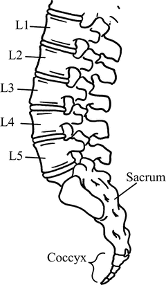
Fig. 7.2
Skeletal anatomy of the lumbosacral spine
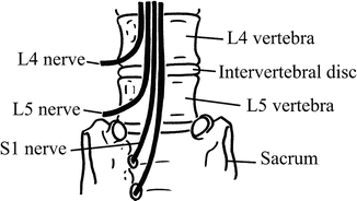
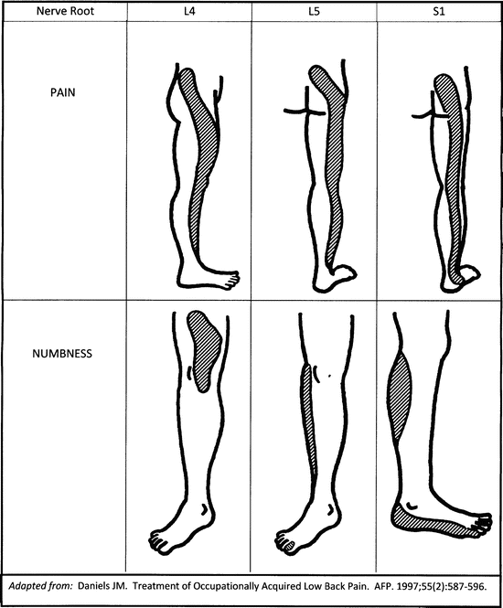
Fig. 7.3
Skeletal and neuroanatomy of the lumbosacral spine at the L4–L5 level
Load in the LS is shared by the lumbar intervertebral disk and the posterior joint facets. The disk itself is made of two sections, the central nucleus pulposus, which is made of a proteinaceous gel, and the outer portion (annulus), which is made of fibrous material. With age, the disks tend to dry out, as there is no direct vascular supply to the disk. This leads to an increasingly friable disk as a person ages, and the nucleus pulposus becomes more solid. When disk herniations occur, they tend to protrude posterolaterally, which then affect the traversing nerve root. This can cause symptoms in the radicular pattern of the nerve involved [3, 4] (Fig. 7.4).
Many theories exist as to the cause of mechanical (nonneurological) back pain, and most patients with mechanical back pain cannot be given a specific diagnosis. Pain may be due to the irritation of muscles, ligaments, or other soft tissues, but a popular theory is that pain is produced by the irritation of the posterior vertebral facets as well as chemicals from the nucleus pulposus [3, 4].
Red Flags
1.
Cauda equina syndrome. This occurs when there is a large herniation or blockage in the lumbar spinal canal and occurs in about 2 % of patients with disk herniations [3]. This is a true surgical emergency, since unless the obstruction is removed quickly (in less than 24–48 h), permanent fecal or urinary incontinence or saddle numbness may occur. It is imperative to inquire about saddle area numbness and incontinence when evaluating a patient with low back pain. When this condition is suspected, CT or MRI and immediate consultation with a spine surgeon are indicated.
2.
History of malignancy or systemic symptoms (weight loss, fever, multiple joint, or multisystem involvement). These patients may have recurrent malignancy or systemic conditions that warrant more aggressive investigation than the average patient with back complaints [4].
3.
Age greater than 50. Patients who present with the low back pain at advanced age warrant also aggressive investigation to rule out nonmechanical causes including cancer. [5]
4.
Progressive neurologic deficit. These patients need more detailed evaluation for their complaints, often involving urgent imaging and/or referral [6].
General Approach to the Patient with Back Pain
Lumbosacral back pain is one of the most common reasons patients visit healthcare providers in the United States [7]. Studies in Canada and Europe have found that the prevalence of low back pain in the general population is 22–48 % [2, 6], and some experts believe that back pain is simply a part of the human condition. In the primary care setting, the physiologic cause of back pain cannot be diagnosed definitively in up to 85 % of patients; however, most patients with back pain will improve within 6–12 weeks. Outcomes are similar when patients see primary care providers, chiropractors, and orthopedic surgeons.
There are three main questions that need to be answered when evaluating the patient with back pain:
1.
Is there evidence of systemic disease?
2.
Is there evidence of neurological compromise?
3.
Are there psychological factors that may contribute to disabling pain?
In the absence of neurologic symptoms or signs, most patients will have mechanical back pain due to benign causes. In general, the history and physical examination is directed at identifying more serious causes that require further investigation.
For all patients, a thorough history should be taken, inquiring about their medical history, medications, presence of any risk factors, trauma, recent infections, and history of prior back injuries, pain, or surgeries. Aggravating and relieving actions should be identified, and if radicular symptoms are present, the pattern can help determine etiology of pain. The red flag symptoms of cauda equina syndrome should always be addressed (see above) [1–3].
A brief vascular examination should always be performed. Palpation of the dorsalis pedis and posterior tibialis pulses is usually adequate. If abnormal, a more detailed examination should be performed.
The back should be inspected for obvious deformity. Palpation of the spine and paraspinous muscles rarely helps make a specific diagnosis; however, isolated vertebral tenderness may indicate bony pathology.
A detailed neurologic exam is not always necessary. All patients should have a straight leg raise test (SLR). This test is done having the patient supine while the examiner passively flexes the hip with the knee extended. A positive test occurs when the patient’s symptoms are reproduced below the knee [1–3]. Reproduction of the patient’s back pain is not a positive test. This test is very sensitive but is not specific; specificity can be improved by addition of the cross-table straight left raise test (described later). Neurological involvement occurs most commonly at the L5 and S1 nerve root levels, and even in the absence of neurologic symptoms, the following assessments should be performed:
1.
Motor L5: have the patient raise (dorsiflex) the great toe
2.
Sensory L5: medial foot and webspace between first and second toes
3.
Motor S1: ankle jerk reflex (may be absent in elderly even without pathology)
4.
Sensory S1: posterior calf and lateral foot
Another option for L5 testing is to have the patient walk on the heels; S1 by walking on the toes. If these examinations are negative in a patient who has no neurological complaints, no further neurological assessment is needed [8].
Related posts:


Stay updated, free articles. Join our Telegram channel

Full access? Get Clinical Tree


