Fig. 5.1
The ankle
The Achilles tendon connects the calf muscles to the calcaneus bone. It is the largest tendon in the body, and tapping this tendon typically results in the ankle jerk reflex. In the event of injury, patients often feel or hear a noise like a loud “pop.” A rupture is usually caused by sudden force being exerted upon the tendon during strenuous physical activity and typically occurs when a patient is pushing off with his/her foot with force. Not all patients feel pain when the tendon ruptures (Gravlee, Hatch, & Galea, 2000). It is important to note that just because a patient retains the ability to plantarflex does not mean that a rupture is not present. Tendon rupture can be effectively diagnosed through clinical examination, but an ultrasound can provide confirmation (Maffulli, 1998).
The ligaments of the ankle are a source of mechanical stability and direct the motion of the joint. Sprains occur when these ligaments tear due to sudden stretching. Often times, ankle sprains are the result of suddenly “twisting” the ankle during sports or stepping off of an uneven surface. These injuries can usually be classified on the basis of physical examination by using the method of injury as a guide to determine the location of the sprain. Inversion and eversion sprains are the two main kinds of ankle sprains. Inversion trauma is responsible for 85 % of ankle sprains (Baumhauer, Alosa, Renström, Trevino, & Beynnon, 1995). This occurs when the foot is twisted inward and the lateral ligaments are stretched too far. Eversion sprains are a result of the foot being twisted outward causing the medial ligament to be stretched too far. Symptoms of a sprain include pain, swelling, and occasionally bruising around the area of injury. A high ankle sprain occurs when the syndesmotic ligament (the ligament above the joint) is injured as well. This kind of sprain can lead to chronic ankle instability (Taylor, Englehardt, & Bassett, 1992). There are three grades of ankle sprains. Grade 1 sprains cause stretching of the ligament, and symptoms are usually limited to pain, tenderness, and swelling. Grade 2 sprains cause a partial tear of the ligaments, and symptoms include pain, swelling, and local hemorrhage resulting in bruising. Patients can usually take a few steps but with considerable pain. Grade 3 sprains result in a complete tear of the ligament(s) and present with significant swelling and the inability to support weight.
An ankle fracture is a common injury that usually has a low complication rate if managed carefully. Nearly five million ankle fractures occur every year in the United States alone (Daly et al., 1987). Some studies have shown a connection between ankle fractures and smoking and high body mass index (Honkanen, Tuppurainen, Kröger, Alhava, & Saarikoski, 1998; Valtola et al., 2002). Clinicians employ the Ottawa ankle rules to help determine whether or not a patient requires a series of X-rays to diagnose a possible bone fracture. The majority of ankle fractures affect the malleolus, which is the bony protrusion on each side of the ankle. These fractures can be broken down into three broad categories: unimalleolar, bimalleolar, and trimalleolar fractures (Court-Brown, McBirnie, & Wilson, 1998; Fallat et al., 1998). An unstable ankle fracture means that there are two or more sites of significant injury. Malleolar fractures tend to be stable if there is no contralateral or syndesmotic injury present. It is important to make sure that these injuries are isolated because medial malleolus fractures can often disturb lateral or posterior structures. Posterior malleolar fractures occur either as part of a pilon fracture or from an eversion force. These kinds of fractures rarely occur in isolation and are usually unstable injuries. Bimalleolar fractures incorporate fractures of both the lateral and medial malleoli. They are unstable and are usually the result of an eversion force. A trimalleolar fracture consists of bimalleolar fracture coupled with a posterior malleolus fracture. These types of fractures are unstable, occur with injuries that involve great force, and have a higher risk of complication.
A pilon fracture occurs when the talus is driven into the articular surface of the tibia by a very strong force. As a result of this force, the distal tibia bones are crushed, and injury is often present in other areas of the body as well. There are two broad categories for pilon fractures depending on the amount of force involved in the accident. The first category is a high-energy injury where the ankle suffers extreme force from something like a car accident. The second category is a low-energy injury where the ankle is compressed due to an activity like skiing.
The human foot is a complex structure that consists of 28 bones. The talus bone joins the foot to the leg. The calcaneus bone forms the heel of the foot and provides the attachment for the Achilles tendon. Plantar fasciitis is one of the most common disorders of the foot and is also known as plantar heel pain. Nearly one million patients complain of this disorder annually in the United States alone (Riddle & Schappert, 2004). Plantar fasciitis is caused by inflammation of the plantar fascia, the thick band of tissue on the bottom of the foot. Plantar fasciitis is usually diagnosed by clinical examination. The most common symptom is pain in the heel or sole of the foot. The pain is usually worse when walking, standing for long periods of time, or after intense physical activity. Possible risk factors may include obesity (Rano, Fallat, & Savoy-Moore, 2001) and reduced ankle dorsiflexion (Riddle, Pulisic, Pidcoe, & Johnson, 2003). Although heel spurs are frequently seen on the X-rays of plantar fasciitis patients (Yi et al., 2011), there is still some debate as to whether there is a correlation between heel spurs and the disorder. The majority of incidents occur in the group of people between the ages of 40 and 60. Plantar heel pain may be the result of other underlying disorders, including atrophy of the heel pad, rupture of plantar fascia, and sarcoidosis (Shaw, Holt, & Stevens, 1988).
Stress fractures are small cracks in a bone that are caused by the repeated application of stress or force. They occur in less than 1 % of the general population (Bennell & Brukner, 1997). Running on a hard surface, repeatedly jumping up and down, or suddenly undertaking an intense workout are examples of some activities that can cause stress fractures. Stress fractures are classified as being at “high risk” or “low risk” of complications depending on the location of the injury. They are usually diagnosed through a physical examination, and early diagnosis is imperative to help avoid further complications. Risk factors include low bone density (Myburgh, Hutchins, Fataar, Hough, & Noakes, 1990), history of a prior stress fracture (Milgrom et al., 1985), increased intensity of physical activity (Mäenpää, Soini, Lehto, & Belt, 2002), low levels of calcium intake, and a low level of physical fitness. Generally, women are at a higher risk for stress fractures than men (Banal et al., 2006). Preventative measures include a well-balanced diet, weight-bearing exercises that improve bone density, and proper training techniques.
Toe fractures are fairly common and account for nearly 9 % of the fractures treated by primary care physicians (Eiff & Saultz, 1993; Hatch & Rosenbaum, 1994). Studies have shown that over 60–75 % of these fractures involve the smaller toes (Schnaue-Constantouris, Birrer, Grisafi, & Dellacorte, 2002). Some common causes of toe fractures include stubbing a toe or a crushing injury caused by a falling object. Less common causes include hyperflexion and hyperextension. Symptoms include swelling, bruising, and throbbing pain. In the case of a displaced fracture, deformity of the toe is usually present. Patients may also complain of difficulty walking or comfortably fitting in shoes.
Occupational Causes of Foot and Ankle Injuries
Several studies have focused on occupational factors associated with the development of lower extremity MSDs. Oftentimes, these studies utilize self-reports to assess musculoskeletal symptoms, without assigning specific diagnoses (i.e., pain, discomfort, or fatigue). When referencing literature on MSDs of the foot or ankle, it is important to note that the lack of significant findings is not the consequence of a demonstrated lack of association, but a result of the sheer deficiency of studies in this area.
One type of foot injury, plantar fasciitis, which is inflammation of the plantar fascia on the bottom of the foot, has been examined within the context of occupational exposure. Investigating plantar fasciitis, Riddle et al. (2003) conducted a study by drawing participants from outpatient clinics. Participants were physician-diagnosed with plantar fasciitis prior to referral, after which time ankle dorsiflexion was measured using a goniometer. Other factors taken into account were time spent on feet and time spent jogging. Using multiple logistic regression modeling, these investigators found a significant association between plantar fasciitis diagnosis and time spent on the feet. However, time spent on the feet was dichotomized (majority or minority of the workday). Additional information about exact duration, time spent walking versus standing, type of surface, and participant footwear was not collected. Consequently, while these researchers present a correlation between standing and plantar fasciitis, conclusions are limited by the workplace factors not accounted for.
Ryan (1989) examined the association between time spent standing and musculoskeletal symptoms of the ankle or foot. Supermarket checkout workers were observed for 10-s intervals every 30 min, after which an activity profile was developed accounting for posture, activity, and department. Compared to other employees, checkout department workers stood the most and had the highest rates of foot and/or ankle symptoms. Although Ryan did not account for BMI, foot and/or ankle symptoms were associated with percent of time spent standing. However, floor surface and shoe design were not described, and these factors are known to affect prevalence of symptoms. Additionally, it should be noted that checkout workers were very young, and the majority were female, limiting the generalizability of these results.
Werner, Gell, Hartigan, Wiggerman, and Keyserling (2010) attempted to overcome the weaknesses seen in some earlier foot and ankle studies by determining the relative influence of floor surface, BMI, age, and work activity in contributing to foot and ankle disorders in assembly plant workers. These researchers also included shoe characteristics and foot biomechanics as independent variables, factors not accounted for in much of the earlier literature. Through self-report questionnaires, 24 % of assembly plant workers were diagnosed with a foot or ankle disorder. Participants with a foot or ankle disorder tended to be older, female, and longer-tenured workers. Previous medical issues such as osteoporosis, rheumatoid arthritis, and heel spurs were also associated with higher prevalence rates of foot or ankle disorders. Overall, results showed an increased risk of foot or ankle disorders associated with more time spent walking, whereby every 10 % of the day spent walking resulted in a 20 % increase in MSD risk. Higher metatarsal pressure was also associated with foot or ankle disorders when adjusted for BMI, time spent standing/walking, and previous medical issue. These results suggest that, when it comes to foot and ankle disorders, there is a complex interplay between a number of intrinsic and occupational factors at work.
The extant research on MSDs of the foot and ankle tend to find a relationship between disorder prevalence rates and time spent standing, higher BMI, and repeated impact (D’Souza et al., 2005). Although limitations exist throughout the literature, there is an undeniable trend across occupations and populations. Generalizability will continue to be an issue due to limitations inherent in this type of research; however, these results serve to influence both primary and secondary strategies for preventing foot and ankle disorders. Continued refinement of epidemiological research into the role of workplace factors in the development of MSDs of the feet and ankle will further clarify specific influences on prevalence rates.
Knee Injuries
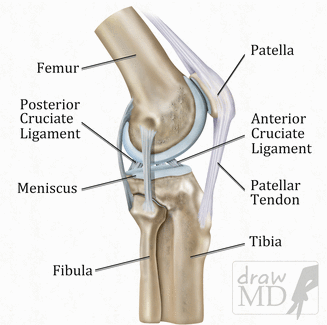
The knee is the largest and most complex joint in the human body. It can be characterized as a modified hinge joint due to the fact that it allows both rotation and flexion while maintaining stability and control under an immense load (Goldblatt & Richmond, 2003). The joint connects the femur to the tibia and fibula through a series of muscles and is stabilized by several thick ligaments. The knee is surrounded by a synovial capsule containing synovial fluid that provides nourishment and lubrication to the joint. Inside the capsule, hyaline cartilage lines both sides of the joint allowing smooth traction. Between the bones lies a fibrocartilaginous c-shaped cushion called the meniscus which provides shock absorption, lubrication, nutrition, stability, and load transmission to the knee joint (Caldwell, Allen, & Fu, 1994). The patella (kneecap) gives significant mechanical leverage to the quadriceps muscles allowing knee extension and straightening as well as a connection between the thigh and shinbones (Fig. 5.2).


Fig. 5.2
The knee
Osteoarthritis (OA) is a common degenerative condition affecting the cartilage in the knee. OA is typically associated with both genetic factors and prolonged mechanical stress (Sandmark, Hogstedt, & Vingard, 2000). Another degenerative condition that affects the knee is bursitis, which is often associated with friction stress causes by repetitive kneeling (Okunribido, 2009).
A torn meniscus is a common injury that frequently occurs when a bent knee is twisted in an unnatural position. Those who participate in physical activity and sports have a high incidence of an acute injury where many sudden movements and cuts are performed. Acute meniscal tears can occur independently. However, the meniscus may also be injured alongside the rupture of a medial collateral ligament (MCL) and the rupture of the anterior cruciate ligament (ACL) (Keene, Bickerstaff, Rae, & Paterson, 1993; Nikolić, 1998). Chronic injury occurs in the elderly where the cartilage of the meniscus is worn down from overuse and degeneration over time. The peripheral (outer) section of meniscus has a higher healing rate, relative to the central meniscus, due to a better vascular supply and subsequently can be repaired (Metcalf & Barrett, 2004). Poor vascularity in the central meniscus requires surgical excision rather than repair (Henning, Lynch, & Clark, 1987).
The ACL is the most common ligament injured in the body that frequently requires surgery (Spindler & Wright, 2008; Vescovi & VanHeest, 2010). The ACL comprises a dense band of connective tissue that connects the femur to the tibia (Dodds & Arnoczky, 1994). The primary function of the ACL is to stabilize the range of motion of the knee by preventing extreme forward translation of the tibia from beneath the femur (Furman, Marshall, & Girgis, 1976). A ruptured ACL occurs when the knee undergoes a large traumatic force in a pivotal motion through both direct and indirect contact (Lin et al., 2012). However, the majority of ACL injuries occur in a noncontact fashion where no physical contact with the knee is involved (Agel, Arendt, & Bershadsky, 2005; Boden, Dean, Feagin, & Garrett, 2000; Feagin & Lambert, 1985). When the ligament is injured, a loss of functional stability occurs, as well as a shearing effect between the femur and tibia due to a lack of constraint by the ACL (DeMorat, Weinhold, Blackburn, Chudik, & Garrett, 2004; Lam et al., 2009; Maffulli, Binfield, & King, 2003). Injuries to the ACL range from micro-tears to complete tears and are classified I to III in severity (Kessler et al., 2008). The majority of ACL injuries are Grade III, where the ligament is completely torn and surgical intervention is required to restore proper function. Participants of high-risk sports account for the majority of torn ACLs where many rapid movements such as deceleration and pivoting take place (Pujol, Blanchi, & Chambat, 2007). Female athletes are more susceptible to an ACL tear than their male counterpart. However, the cause is not completely clear (Carson & Ford, 2011; Cowling & Steele, 2001; Ferrari, Bach, Bush-Joseph, Wang, & Bojchuk, 2001; Hewett, Myer, & Ford, 2001). Injuries to the ACL are also known to increase the risk of knee osteoarthritis occurring later in life (Lohmander, Englund, Dahl, & Roos, 2007; Lohmander, Östenberg, Englund, & Roos, 2004).
Like the ACL, the MCL is important in stabilization and works synergistically with the ACL to stabilize the knee against valgus (bent or twisted outward) stress and tibial translation (Baker & Shalvoy, 1996; Hull, Berns, Varma, & Patterson, 1996). Together, MCL and ACL ruptures account for 90 % of all knee ligament injuries in the young and active (Woo, Chan, & Yamaji, 1997). The typical source of MCL injury is through a direct blow to a bent knee which causes over stretching and subsequent trauma (Gibbs, 1994; Roberts & Stallard, 2000). Sudden pivoting and cutting motions can also indirectly injure the MCL as observed in those that play sports where valgus knee loading is common (Lorentzon, Wedrèn, & Pietilä, 1988; Najibi & Albright, 2005). MCL tears are graded on a scale of I to III in severity, and the general consensus is an MCL injury can be treated in a nonoperative fashion (Fetto & Marshall, 1978; Indelicato, 1983). However, a severe MCL injury may warrant surgical intervention (Edson, 2006; Lonergan & Taylor, 2002; Yoshiya, Kuroda, Mizuno, Yamamoto, & Kurosaka, 2005).
Occupational Causes of Knee Injuries
The literature regarding disorders of the knee tends to focus primarily on osteoarthritis (OA), bursitis, and meniscal lesions/tears. Although knee OA is the most frequently studied disorder of the knee, definitions of OA vary between studies, resulting in some variability across studies (D’Souza et al., 2005). A recent review of research on occupational knee disorders by Reid, Bush, Cummings, McMullin, and Durrani (2010) revealed that the epidemiology of these disorders can be dichotomized into intrinsic (e.g., age, BMI, previous injury) and extrinsic (e.g., occupational) factors. From this, these researchers hypothesized that personal and occupational factors may interact in a complementary manner to predispose individuals to knee disorders. Their review also showed that kneeling is the most often indicated occupational factor identified in knee disorder research and seen to contribute to OA, bursitis, and meniscal lesions. Additionally, squatting and heavy physical work are also often identified as contributing occupational factors to the development of MSDs of the knee.
Knee bursitis presents when inflammation of a bursa sac occurs. This inflammation is often caused by fluid retention, which causes swelling of the bursa sac, and a thickening of the bursa walls (Kivimaki, Riihimaki, & Hanninen, 1994). Knee bursitis is a common outcome in a number of occupations where kneeling or use of the knee as a tool is common (Reid et al., 2010). Using the knee as a tool creates a sudden impact stress, while kneeling for long periods creates a distribution of pressure throughout the knee. For example, carpet layers are noted as uniquely susceptible to knee bursitis as they spend a large amount of time on their knees. Carpet layers also use a tool known as a “knee kicker” for carpet stretching. A number of studies looking specifically at carpet layers have found the force created by knee kickers to be over four times greater than the individual’s body weight (Bhattacharya, Mueller, & Putz-Anderson, 1985; Village, Morrison, & Leyland, 1993). In fact, knee bursitis is so common among carpet layers that it has become known as “carpet layer’s knee” (Kivimaki et al., 1994).
While using the knee as a hammer-type tool is almost entirely unique to the carpet-laying occupation, there are a number of industries where long periods of time are spent kneeling or crawling, including coal mining, plumbing, and assorted carpentry occupations. A number of studies have shown an increase in knee and infrapatellar bursitis as a consequence of time spent kneeling (Partridge, Anderson, McCarthy, & Duthie, 1968; Tanaka et al., 2007; Thun et al., 1987). A review of the literature on occupational knee disorders by Jensen and Eenberg (1996) found an association between time spent kneeling and knee bursitis. While studies have shown a pattern of increased bursitis as a consequence of kneeling work, knee bursitis is a far less serious disorder than knee OA and is therefore not as prevalent in the literature.
Alternatively, knee OA is relatively well represented in the occupational MSD literature. Knee OA affects the cartilage of the knee, causing a loss of cartilage and a narrowing of the joint space. A number of studies have demonstrated a relationship between occupations requiring kneeling or squatting and knee osteoarthritis (Anderson & Felson, 1988; Kivimaki et al., 1994; Tanaka et al., 2007). Kivimaki et al. (1994) looked at the work habits of carpet and floor layers using video recordings while also observing painters as a referent group. Analysis of the recordings revealed that the floor and carpet layers spent 42 % of their workday kneeling on one of both knees, while the painters only spent 5 % of their time kneeling. X-ray examination revealed significantly more osteophytes in the carpet and floor layer group, which are often present in osteoarthritic joints. A case–control study by Cooper, McAlindon, Coggon, Egger, and Dieppe (1994) found that kneeling or squatting for more than 30 min a day significantly increased the risk of knee OA, while Coggon et al. (2000) found that kneeling or squatting for 1 h or getting up from a kneeling or squatting position 30 or more times were significantly associated with knee OA. Additionally, heavy physical work has been associated with knee OA. A study by Lau et al. (2000) matched patients with and without knee OA, confirming OA in diagnosed patients with radiographs. In this study, knee OA was associated with lifting more than 22 lb more than ten times per week in both men and women. Most studies do not list specific weights, but instead delineate workload into light, moderate, and heavy workloads, with definitions for each varying across study. A study by Kohatsu and Schurman (1990) used a self-report questionnaire of workload, finding that participants who reported having worked a job requiring a heavy workload were much more likely to present with knee OA. Anderson and Felson (1988) dichotomized participants by categorizing their occupations into those associated with and without high workload. Their results showed significantly higher prevalence of knee OA in the heavy workload subject group among both men and women. Diagnoses of knee OA were not prevalent among the younger age group, consistent with the development period necessary for chronic OA.
The difference in classifying knee OA between studies may help explain the reported discrepancies in prevalence rates among different studies (Yoshimura et al., 2004). Yoshimura et al. (2004) hypothesize that specific country location may influence the relationship between workplace factors and knee OA. Additionally, Teichtahl, Wluka, and Cicuttini (2003) suggest that future research should attempt to describe the biomechanical factors on knee OA, as well as whether these factors are an influence on risk, or alternatively, are outcomes of occupational factors. Regardless of classification inconsistencies, researchers consistently find a positive relationship between occupational factors, squatting, kneeling, and heavy physical workload and associated knee OA.
Even when compared to research on knee bursitis, the research on meniscal lesions is extremely sparse. A review of these studies by D’Souza et al. (2005) could not support the finding of an association between occupational factors and meniscal lesions. Kirkeskov and Eenberg (1996) point out that very few studies on meniscal lesions of the knee controlled for previous injury, sports participation, or the cause of a traumatic work injury. As such, there is insufficient data to say with confidence that kneeling or squatting influence the development of meniscal lesions. Future occupational studies examining meniscal lesions will need to control for these factors in order to allow for causal inferences.
The literature on knee OA shows a strong association between knee OA and squatting or bending. Knee bursitis has been seen to be caused by both kneeling and squatting across a variety of occupations. There is as yet not enough literature available to draw a conclusion regarding workplace factors influence on meniscal lesions. Generalizing these findings across occupations, locations, and genders is extremely difficult, as workplace biomechanics differ across these factors. Additionally, women are underrepresented in occupations requiring much kneeling or squatting, and men who are currently diagnosed with severe knee OA tend to avoid work requiring them to function in painful positions, potentially confounding prevalence rates. More detailed studies are needed to better understand the relationship between workplace factors and knee OA, bursitis, and meniscal lesions.
Hip Injuries
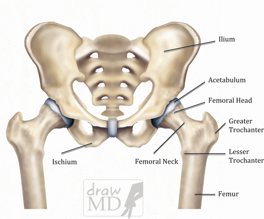
The hip is a ball and socket joint. The round head of the femoral bone articulates with the acetabulum, the cuplike cavity of the pelvis. The femoral head is lined with hyaline cartilage that assists in shock absorption, as well as giving lubricated traction to the joint. Several thick ligaments and the labrum firmly hold the femoral head in the acetabulum. The hip is stabilized by several groups of muscles that allow for a complex range of motions of the hip (Fig. 5.3).


Fig. 5.3
The pelvis
A hip dislocation is a serious and painful injury that results from high-energy trauma. With enough force, the ball-shaped femoral head is displaced from the socket (acetabulum) in the pelvic bone. Dislocation can occur in sports injuries, industrial accidents, and most commonly in motor vehicle accidents (MVA) which account for 62–93 % of all hip dislocation injuries (Sah & Marsh, 2008). In MVA, dislocations commonly result in posterior dislocations of the right hip (Monma & Sugita, 2001; Sah & Marsh, 2008). This is due to the typical position of the driver’s right hip in flexion and adduction. This position effectively increases the chance of a knee-thigh-hip complex impact against the dashboard (Rupp & Schneider, 2004). In a posterior dislocation, the limb is shortened, flexed, and internally rotated whereas in an anterior dislocation, the limb would be flexed externally (Chen et al., 2010). Posterior hip dislocations account for the majority of all hip dislocations, while the anterior dislocation is less commonly observed. Sciatic pain can also manifest as a secondary injury caused by hip dislocation. The sciatic nerve is the longest and thickest nerve in the human body. The nerve originates from the lower spine and runs through the pelvis and down the buttocks into the legs and feet. In a hip dislocation, the peroneal nerve component of the sciatic nerve may be stretched over the displaced femur head, causing pain and numbness (Clegg, Roberts, Greene, & Prather, 2010; Cornwall & Radomisli, 2000; Hillyard & Fox, 2003). The impingement also causes the injured to feel hypersensitive to touch in the lower extremity as well causes an impairment of motor functions (Hillyard & Fox, 2003).
A hip fracture is generally a break in the femoral neck of the femur. The majority of hip fractures occur from a simple fall (as observed in the elderly). However, a small percentage of younger individuals may suffer a hip fracture through a severe impact such as an MVA (Aschkenasy & Rothenhaus, 2006; Cummings, 1996; Keating & Aderinto, 2010; Melton Iii, 1996). The elderly are most susceptible to a hip fracture due to their increased loss of bone density. Furthermore, elderly women with osteoporosis have a higher incidence of fractures than elderly men because of their increased loss of bone mass (Cummings, 1996). Women who smoke also show a tendency to have an increased risk of hip fractures (Cornuz, Feskanich, Willett, & Colditz, 1999; Cummings et al., 1995).
There are three categories of hip fractures: intracapsular, intertrochanteric, and subtrochanteric. Femoral neck fractures (intracapsular fractures) occur inside the joint capsule. This type of fracture may result in partial or complete disunion of the round head from the rest of the femur. The Pipkin classification system is widely used to distinguish the severity of a intracapsular fracture and range from I to IV in severity (Iannacone, Dalsey, & Wood, 1994). In very severe cases, the blood supply may be disrupted, which then may result in avascular necrosis and osteonecrosis of the femur head (Harper, Barnes, & Gregg, 1991; Thuan & Marc). Poor blood circulation to the region creates further complications in healing and frequently leads to nonunion (Roshan & Ram, 2008).
Intertrochanteric (IT) fractures are designated as extracapsular, due to their occurrence outside of the joint capsule. IT fractures occur between the greater and lesser trochanter (large bump of the femur). Several classification systems exist. However, IT fractures are generally classified as either stable or unstable depending on the location, size, and number of bone fragments (Evans, 1949; Jensen, 1980; MacEachern & Heyse-Moore, 1983). A dependable vascular supply allows IT fractures to heal properly. However, other complications may arise. Misaligned healing and a shortening of the femur can occur because of competing forces from the surrounding muscle attachments (Bartonícek, Skála-Rosenbaum, & Douša, 2003; Haidukewych, Israel, & Berry, 2001; Olsson, Ceder, & Hauggaard, 2001).
Subtrochanteric fractures occur between the lower border of the lesser trochanter and 2.5 in. distally in the proximal portion of the femur (de Vries, Kloen, Borens, Marti, & Helfet, 2006; Shukla et al., 2007; Zuckerman, 1996). The subtrochanteric region is subjected to a high level of load-bearing stress and has a poor vascular supply that contributes to slower healing (de Vries et al., 2006; Guerra & Born, 1994). Subtrochanteric fractures are also subject to misalignment and deformities due to the strong forces of the muscular attachments on the femur (Shukla et al., 2007).
Trochanteric bursitis (hip bursitis) is the painful swelling of the bursa that superficially surrounds the trochanter region of the femur and is most commonly presented in the elderly. Many times, this injury is overlooked because of its close nature to other clinical conditions (Mulford, 2007). Micro-trauma from repeated overuse, complications in surgery, direct trauma, as well as a predisposition to other conditions contribute to the development of bursitis (Farmer, Jones, Brownson, Khanuja, & Hungerford, 2010).
Occupational Causes of Hip Injuries
The research on occupational hip disorders has mostly focused on OA of the hip, as well as general hip pain. Relative to research on knee OA, there is relatively little research done on occupational hip OA and even less research on general hip pain. Occupational research on hip OA suggests a relationship between development of hip OA and workplace biomechanical factors. In a review of the hip OA literature, Maetzel, Makela, Hawker, and Bombadier (1997) found an association between certain work factors and hip OA in men. This review did not include any studies examining hip OA in women. A more recent review by Schouten, de Bie, and Swain (2002) examined previous studies with strong methodology, finding a significant relationship between the development of hip OA and heavy lifting in both men and women. More recent studies have found correlations between heavy lifting exposure and hip OA, although what is considered “heavy lifting” varies across studies, and some studies simply extrapolate expected lifting exposure using only job title (D’Souza et al., 2005).
Hip pain has also been the focus of a small number of occupational studies. However, these studies tend to be extremely limited in their scope and often have a number of methodological flaws. Similar to the literature on the ankle and feet, the literature on the influence of work factors on hip disorders is extremely limited. Although patterns have emerged linking hip OA to certain workplace factors, further research is needed to fully understand the relationship between these factors and the development of MSDs of the hip.
Stay updated, free articles. Join our Telegram channel

Full access? Get Clinical Tree


