Abstract
Objectives
Back pain is often attributed to increased tension in the back muscles, regardless of whether the tension is primary or related to a disc/facet pathology. We hypothesized that when either lower back pain or neck pain is unilateral, the muscle tension would be more pronounced on the painful side and could be detected by palpation alone (i.e. without the need to apply pain-triggering manoeuvres).
Methods
Patients with unilateral neck or lower back pain were enrolled in the study. Participants with scoliosis, obesity, a history of spinal surgery or pain radiating below the knee or the elbow were excluded. The patients were examined by comparative muscle palpation only. The examiner was unaware of which body side was painful and the patient was told to remain still and silent. The spinal muscles were examined bilaterally, with superficial and deep palpation. The examiner had to determine on which side the tension was greater. The patients’ age, body mass index, time since onset of symptoms and Rolland Morris (lower back pain) and INDIC (neck pain) functional disability questionnaire scores were recorded.
Results
Ninety-one patients with unilateral lower back pain (35 males, 56 females; mean ± SD age: 45.2 ± 15 yrs) and 94 patients with unilateral neck pain (26 males, 68 females, 49.1 ± 15 yrs) were enrolled in the study. The lower back pain and neck pain were right-sided in 50 (54.9%) and 53 (56.4%) of cases, respectively. The examiners correctly identified the painful side in 64.8% of the cases of lower back pain (a significantly better percentage than chance alone (i.e. 50%), P = 0.02) and 58.5% ( P = 0.10) of the cases of neck pain. In lower back pain patients, the results were better for right-side pain than for left-side pain (70.0% and 58.5% of correct answers, respectively, ns). In neck pain patients, the results were better for left-side pain than right-side pain (61% and 56.6%, respectively, ns). There were no significant differences between the two examiners’ respective performance levels. The patients’ clinical parameters did not appear to influence successful detection of the painful side.
Conclusion
Our findings suggest that palpation can detect increased muscle tension in a limited proportion of cases.
Résumé
Objectifs
La douleur de dos est souvent attribuée à une tension augmentée des muscles du dos, quelle qu’en soit la cause (primitive ou secondaire à une pathologie discale ou facettaire). Nous avons fait l’hypothèse qu’en cas de douleur unilatérale (lombaire ou cervicale), la tension musculaire devrait être plus marquée du côté douloureux et qu’elle pourrait être détectée par la palpation seule, à l’exclusion des manœuvres de reproduction de la douleur.
Méthode
Les patients avec une douleur lombaire ou cervicale unilatérale furent enrôlés. Furent exclus les patients avec scoliose, obésité, antécédents de chirurgie vertébrale ou douleur irradiant au-delà du genou ou du coude. Les patients ne furent examinés que par palpation musculaire comparative. L’examinateur ignorait le côté douloureux, et le patient devait rester passif et muet. Les muscles spinaux étaient palpés des deux côtés (palpation superficielle et profonde). L’examinateur devait déterminer de quel côté la tension était la plus marquée. L’âge, l’IMC, la durée des symptômes et les résultats des questionnaires EIFEL et INDIC furent notés.
Résultats
Quatre-vingt-onze patients avec lombalgie unilatérale (35 hommes, 56 femmes, âge 45,2 ± 15) et 94 avec cervicalgie unilatérale (26 hommes, 68 femmes, âge 49,1 ± 15) furent disponibles pour l’analyse finale. La douleur était à droite dans respectivement 50 (54,9 %) et 53 (56,4 %) cas. Le côté correct fut trouvé dans respectivement 64,8 % des cas, un résultat meilleur que le hasard (50 %, p = 0,02) et 58,5 %, un résultat non différent du hasard ( p = 0,10). Chez les patients lombalgiques, les résultats furent meilleurs pour le côté droit que pour le gauche (70,0 % contre 58,5 % de bonnes réponses, ns). Chez les patients cervicalgiques, ils furent meilleurs pour le côté gauche (61 % contre 56,6 %, ns). Il n’y eu pas de différence significative entre les deux examinateurs. Il n’y avait aucune influence des différents paramètres cliniques sur les taux de succès.
Conclusion
Notre étude montre que la palpation peut reconnaître une tension musculaire augmentée dans un nombre limité de cas.
1
English version
1.1
Introduction
One of the cornerstones of manual medicine is that changes in soft-tissue texture revealed by palpation have diagnostic value, independently of pain triggering . These changes in texture concern mainly the paraspinal muscles, which are reportedly the site at which tension (also referred to as “muscle spasm” or “muscle contraction”) is elevated in patients suffering from back or neck pain . In manual medicine textbooks, this increase in tension is described as greater hardness of the muscle body on palpation. It may accompany anatomic or functional damage to the motion segment and may in turn be a potential source of pain . However, this hypothesis has been strongly criticized by other researchers. According to Bogduk, “the entity of « muscle spasm » has no validity, for there is no known neurophysiological correlate of this clinical sign. Moreover, the reliability of finding muscle spasm has been so poor as to defy reporting in terms of kappa scores” .
This context prompted us to adopt another research strategy by looking for muscle tension in patients with unilateral neck or lower back pain.
1.2
Patients and methods
Our working hypotheses were that:
- •
when the neck or lower back pain was unilateral, it would be accompanied by increased muscle tension on the same body side and;
- •
this muscle increase in tension could be detected manually.
Our protocol was simple: patients with unilateral neck or lower back pain were examined by an examiner who had to determine the painful side by muscle palpation alone and without seeking to trigger pain. The examiner was blinded to the patients’ pain status (i.e. painful side). If the examiner could do better than chance, our two hypotheses would be true. If the opposite was true, one or other or both of our hypotheses would be false.
1.2.1
The clinical examination procedure
Two physicians with training in manual medicine participated in this study. Patients were recruited from our university hospital’s back pain clinic and had to have unilateral neck or lower back pain (whether acute or chronic). The unilateral nature of the pain was confirmed by the patient himself/herself, who was asked to indicate the body side and the painful with his/her finger. Patients in whom pain radiated beyond the knee (for lower back pain) or the elbow (for neck pain) were not included. The other exclusion criteria were as follows: symptomatic lower back pain or neck pain, the presence of scoliosis, a history of vertebral surgery, a body mass index (BMI) over 30 or poor understanding of the French language. Once the patient enrolled, the examiner was called in. The examiner palpating the patient was not aware of which side was painful. The examination consisted solely of comparatively palpating the muscles in the painful area. The patient was asked to remain still and silent and, in particular, to refrain from pain-avoidance reactions or facial reactions or verbal complaints if he/she felt pain. The other investigator checked that this rule was applied. In the event of non-compliance, the patient was eliminated from the study. The palpated muscles were as follows:
- •
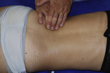
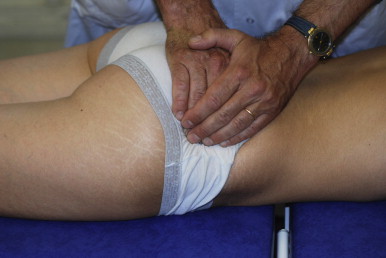
the paraspinal muscles (the multifidus and spinal erectors) and the buttock muscles in cases of lower back pain, with the patient in the prone position ( Figs. 1 and 2 ) and;

Fig. 1
Palpation of the lower back paraspinal muscles, with the subject in the prone position. Examination of the multifidus appeared to be the most informative procedure.

Fig. 2
Palpation of the buttock muscles.
- •
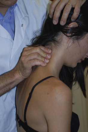
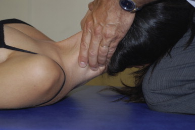
the rear neck muscles, the trapezius muscles and the levator scapula in cases of neck pain, with the patient sitting down and then in the supine position ( Figs. 3 and 4 ).

Fig. 3
Palpation of the rear neck muscles in the sitting position, with the head slightly tilted forwards.

Fig. 4
Palpation of the rear neck muscles, with the subject in the supine position.
The objective was to evaluate the muscles’ texture and determine on which side they were firmest. No other examination manoeuvres were allowed (notably assessments of overall or segmental spinal mobility or a straight-leg raise). The examination’s outcome was binary (i.e. higher tension on the right or higher tension on the left) for the overall muscle status and not muscle by muscle. This outcome was then compared with the truly painful side and was classified as correct (i.e. the painful side was correctly identified) or incorrect. An impression of symmetry (i.e. the same tension on the right and the left) was also considered to be an incorrect answer.
In order to determine factors that could be potentially predictive of a correct answer, we asked the patients to fill out a self-questionnaire with the age, BMI, an assessment of pain during the consultation on a 10-cm visual analogue scale (VAS) and the EIFEL functional disability score (a French-language version of the Roland-Morris Disability questionnaire) for cases of lower back pain or the Indice de Douleur et d’Incapacité Cervicale (INDIC) scale functional disability score for cases of neck pain. We decided to include 100 patients with lower back pain and 100 with neck pain. Each of the two participating physicians had to examine 50 patients from each group. In the absence of any studies of this type in the literature on which to base our sample size calculation, we sought to make our study as powerful as possible by including as many patients as possible. A pilot study with 15 patients enabled us to harmonize the examiners’ clinical examination procedures.
1.2.2
Statistics
The data are presented as the mean average (± 1 standard deviation). To determine each examiner’s ability to identify the painful side correctly, we compared the observed success rate with the theoretical percentage that would be obtained by chance alone (i.e. 50%). To determine the predictive value of a positive result, the data were compared with a Student’s t test (for quantitative variables) or a Chi 2 test (for categorical variables). The threshold for statistical significance was set to P < 0.05.
1.3
Results
Of the 200 enrolled participants, nine patients with lower back pain and six patients with neck pain were eliminated from the study due to uncontrolled pain avoidance (even for mild pain) or verbal complaints that invalidated the blinded nature of the palpation. Hence, 91 patients with lower back pain (39 with acute pain and 52 with chronic pain) and 94 patients with neck pain (35 with acute pain and 59 with chronic pain) were available for analysis ( Table 1 ).
| Male/female gender ratio | Age (years) | Median time since onset of the pain (months) | Right/left side | |
|---|---|---|---|---|
| Lower back pain n = 91 | 35/56 | 45 ± 15 | 6 | 50/41 |
| Neck pain n = 94 | 26/68 | 49 ± 15 | 5 | 53/41 |
1.3.1
Lower back pain
The correct side was identified in 59 of the 91 cases (64.8%), i.e. a significantly better result than chance alone (50%; P = 0.02). Table 2 presents the results for each body side in detail. Although the success rate was slightly higher on the right side than on the left side, this difference was not statistically significant. We did not observe any influence of the various clinical parameters on the success rate ( Table 3 ).
| True side | Correct answers | Incorrect answers |
|---|---|---|
| Right lower back pain n = 50 | 35 (70%) | 15 |
| Left lower back pain n = 41 | 24 (58.5%) | 17 |
| Correct answers | Incorrect answers | P | |
|---|---|---|---|
| Age | 44.1 ± 16 | 47.1 ± 14 | 0.37 |
| BMI | 23.8 ± 3.4 | 23.2 ± 5.4 | 0.99 |
| VAS pain out of 10 | 4.6 ± 2.2 | 4.3 ± 2.2 | 0.54 |
| EIFEL score | 8.3 | 7.5 | 0.40 |
| Male gender | 67.6 % | 32.4 % | 0.99 |
1.3.2
Neck pain
The correct side was identified in 55 of the 94 cases (58.5%); the difference with respect to a value of 50% that would have been obtained by chance alone was not statistically significant ( P = 0.10). Table 4 presents the results for each body side. Although the success rate was slightly higher on the left side than the right side, this difference was not statistically significant. We did not observe any influence of the various clinical parameters on the success rate ( Table 5 ).


