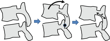
Fig. 31.1
Generational characteristics of LBP
The pathomechanisms of LBP vary according to age. Sports-related stress on immature spinal vertebrae during the growth phase can cause end-plate disorders, and stress fractures of the vertebral arch (spondylolysis). Intervertebral disc degeneration begins in youth and adulthood, and minute damage to intervertebral discs can allow nerve tissue to growth into the annulus fibrosus, causing intervertebral discogenic pain. Discogenic LBP can also be brought about by mechanical weakness that causes the nucleus pulposus to puncture the annulus fibrosus. Further, a simultaneous reduction in intervertebral disc height can decrease disc load functions, which increases stress on the facet joints and can easily cause facet joint pain. Degenerative changes progress with age, leading to osteoarthritis of the spine and stenosis of the spinal canal.
31.2.1 Lumbar Intervertebral Disc Disorders/Disc Herniation
Reduced levels of proteoglycans, which generally occurs during the aging process, decrease water content in the nucleus pulposus, causing further degeneration of the lumbar intervertebral discs. Several studies are currently investigating the risk factors associated with intervertebral disc degeneration that accompanies spinal degeneration. The results of an epidemiological survey we conducted on middle-aged and elderly subjects showed that a history of sports activity is a risk factor, in addition to advanced age, high BMI, high levels of low-density lipoprotein (LDL) cholesterol, and heavy labor (Hangai et al. 2008). We also conducted a cross-sectional survey comparing lumbar intervertebral disc degeneration rates among members of sports clubs at a physical education university and subjects with no sports history to clarify the relationship between sports activity and intervertebral disc degeneration. The results showed significantly higher rates of disc degeneration in baseball players (60 %) and competitive swimmers (58 %) compared to the control group (31 %) (Hangai et al. 2009). Comparing LBP severity with the presence of intervertebral disc degeneration showed higher rates of disc degeneration as LBP severity increased. Several previous surveys have failed to clarify the relationship between LBP and lumbar intervertebral disc degeneration. However, such a relationship has been suggested in subjects with ages close to or around 20 years.
We are carrying out a continuous cross-sectional survey on intervertebral disc degeneration based on the type of sport. The results have shown high rates of disc degeneration in volleyball players (69 %), weightlifters (62 %), and rowers (60 %). These rate differences in disc degeneration among sports led us to believe that the specific physical actions of each sport apply stress to the intervertebral discs, which contribute to degeneration.
Intervertebral disc herniation is broadly divided into bulging type, subligamentous protrusion type, transligamentous protrusion type (protrusion into the spinal canal through the posterior longitudinal ligament), and sequestration type, with each type having a different natural process, prognosis, and treatment. Further, in cases of congenital spinal canal stenosis, even small herniated discs can compress the nerve root between the herniation and the vertebral arch. In these cases, even if the severe early symptoms of nerve root stimulation improve, later stimulation such as through exercise can cause continuous LBP and leg pain. Such compression-type nerve root disorders are difficult to treat and many require surgery. The Kemp test often generates positive results in these cases.
31.2.2 Extension Type Low Back Disorders
Lumbar vertebral arch stress fracture (lumbar spondylolysis), facet joint disorders, and repeated extension or rotation of the lower back and trunk focuses stress on the area between the articular process of the vertebral arch (Fig. 31.2), which can cause stress fractures in this area during the growth stage (Terai et al. 2010) After the bones mature, this kind of stress on the facet joint can lead to disorders involving this joint that present with pain when extension of the lumbar vertebrae is restricted and at the end of extension, as well as pressure pain on the spinous processes of the affected vertebrae. LBP can also be induced by restricting extension and rotation of the lumbar vertebrae (Kemp maneuver) (Note: When there is nerve root compression due to spinal canal stenosis in the lower back, this maneuver will induce pain in the legs and the result of the Kemp test will be positive). Because the nerve root runs ventrally through the area where the vertebral arch separates, growth of fibrous cartilage in this area can put pressure on the nerve root, inflammation of the facet joint, or area of separation can spread to the nerve root, and any of the above can cause leg pain or numbness. Lumbar spondylolysis is classified into three stages: early stage, progressive stage, and terminal stage (Fig. 31.3). Because it is difficult to render the area of separation using simple radiographic oblique views in the early and progressive stages, CT imaging is needed for early diagnosis. Synostosis can be achieved through conservative treatments in the early and progressive stages, in which high intensity changes which indicates bone edema are visible in the interarticular portion of the vertebral arch in magnetic resonance imaging (MRI). Orthotic treatments that restrict extension and rotation of the trunk are used in such cases. However, conservative treatments are not likely to achieve synostosis in terminal stage cases that exhibit clear signs of pseudoarthrosis in simple X-ray images. Thus, treatment of terminal stage lumbar spondylolysis focuses on LBP countermeasures such as stretching the muscle groups of the trunk and legs, and training the muscles of the trunk. Even in cases where separation cannot be confirmed in image findings, cases with similar spinal findings should be considered as a preliminary stage (early stage spondylolysis) .


