Fig. 8.1
Varus osteoarthritic knee with tibial metaphyseal deformity, (a) preoperative radiograph, (b) postoperative radiograph following single stage TKA
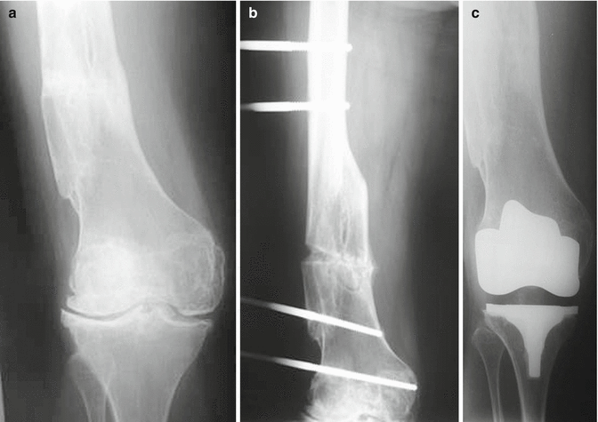
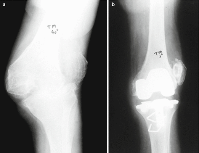
Fig. 8.2
Valgus osteoarthtitic knee with femoral diaphyseal deformity, (a) preoperative radiograph, (b) radiograph showing extraarticular corrective osteotomy, (c) postoperative radiograph following staged TKA
In this review, the effect of the severity of the arthritic knee deformity on the long term performance of TKA is evaluated.
Knee Joint Deformity
Malalignment in the frontal plane is a common manifestation of knee arthritis, and it remains uncertain whether it is a cause or a consequence of the degenerative pathway [6]. Knee joint alignment, as determined by the mechanical axis, can be measured on long full limb radiographs as the angle formed by the intersection of the line connecting the centers of the femoral head and intercondylar notch (femoral mechanical axis) with the line connecting the centers of tibial spines and the ankle talus (tibial mechanical axis) [7, 8]. In the neutral knee, the two axes should be collinear, forming the load bearing axis, where the knee center should be located [9]. A knee can be described as being varus when alignment is >0° in the varus direction, and valgus when alignment is >0° in the valgus direction [7]. An alternative way of indicating varus and valgus malalignment on radiographic imaging is using angle values <180° and >180° respectively [6]. Although the use of the mechanical axis is considered the gold standard, alignment on the frontal plane can be safely determined by the anatomical axis using a pre-existing plain radiograph of the knee, or by certain clinical measures, avoiding unnecessary radiation exposure and facilitating assessment for researchers and clinicians [6]. Increased valgus deformity poses the risk of patellofemoral osteoarthritis progression [10]. Fixed flexion deformity is another common angular deformity seen in knees with indications for TKA. The lack of full knee extension results in greater quadriceps contraction, altered gait kinematics and overloading of the contralateral limb [11].
Varus Deformity
Varus deformity is the most common angular contraction of the knee joint indicated for primary TKA [6, 12] (Fig. 8.3). Arthritic knees with varus deformity are characterized by cartilage and/or bone loss (mainly tibia) in the medial compartment. As a result of the angulation, soft tissues and ligaments of the medial side undergo contracture and must be released in order to achieve neutral limb alignment in TKA [12]. Concerning the definition of severe varus deformity, there is a lack of consistency within the available literature. Some authors define severely varus deformed arthritic knees as bearing a femoral mechanical axis/tibial mechanical axis angle of 8° or at least 10–12° in varus [8, 13, 14]. Other studies use angles of >15° or 20° [15–18]. These variations possibly reflect the authors’ decision to divide the available patient population into subgroups, in order to make assumptions related to the preoperative joint deformity. Angulations of more than 20° around the joint metaphysis usually require a procedure additional to TKA to efficiently restore limb alignment [4]. According to Engh [5], the choice of ligament release technique depends on the severity of varus deformity. Almost all surgeons are familiar with soft tissue release of the medial side of the proximal tibia. Joint line release with sub periosteal fractional detachment of the superficial and deep medial collateral ligaments has proven successful for mild to moderate varus deformity. When it comes to severe varus deformity with fixed flexion contraction, an epicondylar osteotomy to release the collateral ligaments from the femur is useful. Thus, not only is balancing of the varus deformity achieved, but also better access to the posterior contracted structures of the knee. Posterior cruciate ligament and sometimes the popliteus tendon are the causes of these combined contractures. In such cases, balancing of the posterior cruciate ligament or sacrificing it and the use of a more constrained implant are commonly used options [5]. Extensive medial release can inevitably result in increased medial flexion gap and the mandatory use of a thicker polyethylene insert or a more constraint prosthesis [12].
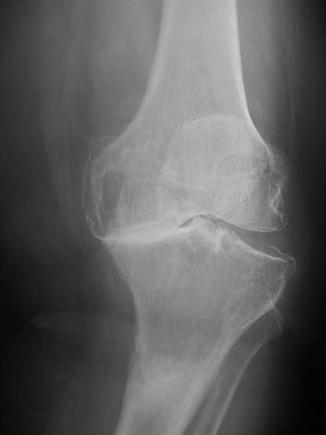
Fig. 8.3
Radiograph showing severe varus deformity of a left osteoarthritic knee
Surgical Technique
Various surgical techniques have been used in order to successfully reconstruct severe deformities during TKA. Mullaji et al. [19] used medial extra articular tibial osteotomy combined with selective posteromedial release. In all cases posterior cruciate substituting implants were used with tibial stem extenders and bone grafting, when necessary, based on the size of tibial bone defects. These authors report very good mid- term results regarding stability and deformity correction [19]. Meftah et al. [20] proposed a posteromedial capsulotomy, piecrust incising of the superficial medial collateral ligament and manipulations under valgus stress, in an attempt to minimize complications derived from extensive medial release. Satisfactory mid-term results for severe varus and fixed flexion were reported in terms of deformity correction, reduced incidence of over release and instability, hematoma formation, and the need for constrained prosthesis [20]. Dixon et al. [18], in a small series of patients, proposed a technique of downsizing, lateralizing of the tibial component and removing the excess proximal medial tibial bone in order to achieve loosening of the medial side and correction of severe varus deformity. In the most cases, non-constraining implants were used. The authors claim excellent clinical and radiologic results and stable correction in mid-terms [18]. According to their opinion, longer term data is necessary in order to identify possible increased polyethylene wear due to tibial prosthesis lateralization. Mullaji et al. [21] described a sliding medial condylar osteotomy performed under navigation guidance. Correct repositioning of the condylar fragment and knee ligament balancing was optimized by computer assisted navigation and the authors reported satisfactory mid-term results [21].
Few long term clinical outcome studies are available. Ritter et al. [22] suggested that patients with severe deformity should not be excluded from surgical treatment having as sole criterion the deformity itself. They report mid to long term results with no significant difference in terms of function, alignment or implant failure rates compared to a control group without severe deformity [22]. In all cases, different types of posterior cruciate ligament retaining prostheses were used. In their opinion constrained implants can be used in extreme cases. Kharbanda et al. [15] evaluated the use of grafting techniques for the reconstruction of bone defects in primary TKA for severe varus knees and identified the indications for structural or impaction grafting, depending on the extent of bone defect. Long term outcome was excellent, justifying the use of such cost effective, biological agents. Implants with stem extenders were used in almost all cases [15]. Karachalios et al. [23] studied the clinical outcome of severe varus and valgus arthritic knees reconstructed with the use of posterior cruciate retaining prosthesis. Outcomes of the varus subgroup were comparable to those of arthritic knees without severe deformity.
The hypothesis that computer assisted surgery (CAS) can efficiently contribute to the satisfactory reconstruction of severe knee deformities in TKA has been recently tested (Fig. 8.4). Several authors have attempted to correlate navigation guided TKA to better early postoperative results but failed to provide longer term superiority. Hsu et al. [14] supported the accuracy of CAS reconstruction despite the fact that severe deformity negatively affected the postoperative axis. Bae et al. [17] positively correlated the severity of varus deformity with postoperative alignment, while Maniwa et al. [16] found no such correlation or higher rate of complications. Moon et al. [12] suggested that the preoperative degree of varus deformity and proximal tibia vara, among other clinical and radiological parameters, were the only determinants of the accuracy of varus deformity reconstruction. In a meta-analysis of twenty nine studies, results favored the use of computer navigated compared to conventional TKA, without taking into consideration the severity of deformity [24].
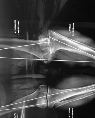
Fig. 8.4
Complex combined femoral and tibial deformity. Indications for computer assisted surgery
Valgus Deformity
Arthroplasty in the valgus knee is a challenging procedure not only for the surgeon, but also in terms of instrumentation [25] (Fig. 8.5). It should not be considered the reverse of varus deformity, as it is different and far more demanding [5, 26] (Fig. 8.6). Distal femoral anatomy is often distorted with the lateral femoral condyle abnormally small, almost dysplastic. Lateral ligaments are contracted and medial soft tissue structures stretched. Required asymmetric resections of the femoral condyles for limb alignment can cause further ligamentous imbalance [5]. Thus soft tissue balanced release becomes a crucial element for satisfactory TKA outcome [25, 27, 28]. Appropriate limb alignment should be obtained intraoperatively by symmetrical flexion and extension gaps and a centralized patella position [25, 29]. Deformities of the adjacent joints may be also present and should be addressed. Valgus flat foot is often associated with valgus knee deformity. The surgeon can consider correction of these deformities prior to TKA in order to ensure proper alignment and function [26].
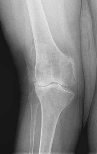
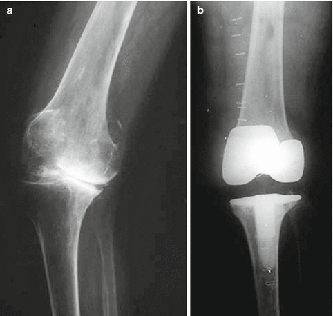
Fig. 8.5
Radiograph showing severe valgus deformity of a right osteoarthritic knee
Fig. 8.6
Severe valgus deformity of a right osteoarthritic knee, (a) preoperative radiograph, (b) postoperative radiograph showing satisfactory limp alignment
Valgus deformity greater than or equal to 20° is considered severe by some authors [22, 30], while others accept smaller angles (10–15°) as the lower limit to severe deformity [28, 29, 31]. Ligament structure parameters such as medial collateral ligament insufficiency and the potential of posterior cruciate ligament sacrifice, favoring the use of a constrained prosthesis, should be taken into consideration [30]. Several authors claim higher failure rates and difficulties in revision of constrained prostheses and they suggest the use of these implants in the elderly and less demanding patients [32]. Peroneal nerve palsy can occur when treating knees with severe valgus deformity. When knee alignment is restored, the nerve is stretched. Spinal or epidural anesthesia may induce this complication and hide its symptoms [26]. Rajgopal et al. [30] have suggested that the operated knees should be placed in 10° of flexion during the early postoperative period in order to avoid peroneal nerve elongation [30].
All authors highlight the importance of proper lateral soft tissue release. Ritter et al. [22] noted that surgeons tend to slightly undercorrect severe deformities. Koskinen et al. [33] suggested that residual valgus deformity increases the risk of revision and all patients with preoperatively severe valgus deformity should be followed up regularly, especially for implant wear and late onset instability. Various authors report satisfactory long term outcomes using cruciate retaining implants, in terms of alignment, revision and delayed instability [22, 27, 30]. Politi et al. [29] used a lateral cruciform retinacular release in combination with posterior cruciate retaining implants and demonstrated satisfactory mid to long term results. Nikolopoulos et al. [31] used a lateral parapatellar approach and tibial tubercle osteotomy instead of the standard medial parapatellar approach and reported satisfactory functional long term results. Radulescu et al. [26] do not favor the lateral approach due to soft tissue complications and inappropriate wound coverage after deformity correction. Zhou et al. [34] favor the lateral approach but also suggest that medial collateral ligament reconstruction and tibial tubercle osteotomy have a high complication rate and increased surgery time. Augmentation of the deficient lateral condyle is an option but the indications and results of this technique are inconsistent in the literature [33, 34]. Ranawat et al. [28] have described a combination of inside-out release technique which involves the capsule, the iliotibial band, appropriate bone cuts and the use of posterior stabilized implants, and reported satisfactory long term outcomes. Marked valgus deformity which requires extensive posterolateral release in combination with medial collateral ligament insufficiency undermines the stability of the arthroplasty in the long term. Knees with such an intraoperative ligamentous imbalance should be replaced with a more constrained implant in order to ensure satisfactory long term results [22, 30, 33]. Zhou et al. [34] treated a series of severe valgus knees with marked osseous deficiency using different techniques and implants (cruciate-retaining, posterior stabilized or hinged knees), individualizing the decision based on the degree of soft tissue contraction, bone loss and intraoperative stability. They reported excellent long term results, supporting the use of the less possible constrained prostheses, especially in cases of younger patients. In their opinion, having in mind a future revision, preservation of bone stock offers satisfactory long term outcome. Finally, Karachalios et al. [23] studied the clinical outcome of severe varus and valgus arthritic knees reconstructed with the use of posterior cruciate retaining prostheses. Outcomes of the valgus subgroup were also comparable to those of arthritic knees without severe deformity. However, in valgus deformities, when exceeding 30°, a high incidence of extensor mechanism complications were observed (Fig. 8.7).
 The Long Term Outcome of Total Knee Arthroplasty. The Effect of Age and Diagnosis
A Brief History of Total Knee Arthroplasty
The Long Term Outcome of Total Knee Arthroplasty. The Effect of Age and Diagnosis
A Brief History of Total Knee Arthroplasty
 Long Term Survival of Total Knee Arthroplasty. Lessons Learned from the Clinical Outcome of Old Designs
 Long Term Outcome of Total Knee Arthroplasty. The Effect of Implant Fixation (Cementless)
 Long Term Outcome of Primary Total Knee Arthroplasty. The Effect of Body Weight and Level of Activity
 Mid to Long Term Clinical Outcome of Medial Pivot Designs


Related posts:
The Long Term Outcome of Total Knee Arthroplasty. The Effect of Age and Diagnosis
A Brief History of Total Knee Arthroplasty
Long Term Survival of Total Knee Arthroplasty. Lessons Learned from the Clinical Outcome of Old Designs
Long Term Outcome of Total Knee Arthroplasty. The Effect of Implant Fixation (Cementless)
Long Term Outcome of Primary Total Knee Arthroplasty. The Effect of Body Weight and Level of Activity
Mid to Long Term Clinical Outcome of Medial Pivot Designs
Stay updated, free articles. Join our Telegram channel
Full access? Get Clinical Tree
