Sterile Instruments/Equipment
- Dental picks
- Freer and AO elevators
- Spiked pusher
- Kirschner wires (0.35, 0.45, 0.54, and 0.62 mm)
- Small point-to-point bone clamps
- 2.0- and 2.4-mm screws
- 3.5- and 4.0-mm cortical screws
- 2.4- and 2.7-mm plate/screw sets (straight and T-shaped)
Surgical Approaches
Dorsal Approach to the Midfoot
- Patient positioning
- Supine.
- Radiolucent table of cantilever type.
- Bring the patient to cantilever (foot) end of the table.
- Place a bump beneath the ipsilateral buttock and flank to neutralize limb rotation.
- Prep and drape the affected lower extremity to the ipsilateral groin.
- Place the appropriately sized sterile radiolucent triangle under the knee.
- Supine.
- Fluoroscopically, locate the first intermetatarsal space on the AP view.
- Incise the skin and subcutaneous tissue longitudinally directly over the first intermetatarsal space.
- Take care to preserve the branches of the superficial and deep peroneal nerves and the dorsalis pedis artery.
- Retract medially or laterally depending upon their position.
- Take care to preserve the branches of the superficial and deep peroneal nerves and the dorsalis pedis artery.
- Dissect the capsules overlying the first and second metatarsocuneiform joints.
- Typically, these capsules will have been disrupted traumatically.
- Incise the joint capsule along the articular borders of the first metatarsocuneiform articulation to visualize the articular surfaces dorsally, medially, and plantarly.
- Incise the joint capsule along the articular borders of the second metatarsocuneiform articulation to visualize the articular surface dorsally, dorsomedially, and dorsolaterally.
- When the third, fourth, and/or fifth metatarsocuneiform joints are subluxated, dislocated, or fractured, a second incision should be made over the interspace between the third and fourth metatarsals.
- Adequate visualization of the third metatarsocuneiform joint is difficult through an incision placed over the first intermetatarsal space, requiring the use of this second incision.
- Determine the location of this incision by identifying the metatarsocuneiform joints and the interspace between the third and fourth metatarsals fluoroscopically.
- Incise the skin longitudinally directly over the interspace between the third and fourth metatarsals and extend this incision proximally, dissecting through skin and subcutaneous tissue.
- Dissect to bone, retracting tendons and neurovascular structures (medially or laterally, as appropriate) to visualize the third and fourth metatarsocuneiform joints.
- Adequate visualization of the third metatarsocuneiform joint is difficult through an incision placed over the first intermetatarsal space, requiring the use of this second incision.
Crush/Open Injuries
- Consider the use of spanning external fixation to temporize and provisionally stabilize the bony and/or ligamentous midfoot injury.
- Definitive open reduction and internal fixation should be performed once edema has subsided and the soft tissue injury has healed sufficiently to allow a safe surgical approach.
- Soft tissues must be followed closely for 1 to 6 weeks in order to determine the time at which incisions for open reduction can be made so as to minimize associated complications.
- Definitive open reduction and internal fixation should be performed once edema has subsided and the soft tissue injury has healed sufficiently to allow a safe surgical approach.
- Severe crush or open injuries, particularly plantar degloving “slipper foot” injuries and extensive plantar lacerations, should be considered for amputation (Fig. 25-1).
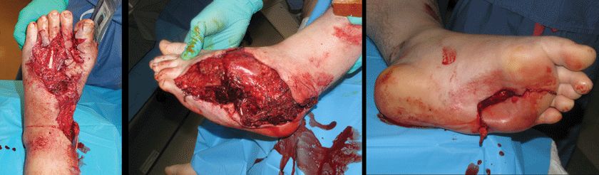
Figure 25-1. Examples of severe mangled foot injuries that should be considered for amputation.
Reduction and Implant Techniques
External Fixation
- Recommended for initial (provisional) treatment of severe crush or open injuries of the foot, especially in the presence of instability, shortening, dislocation, or deformity.
- Typically, this first treatment stage is converted to internal fixation when the condition of the soft tissues allows a safe surgical approach.
- May also be used to supplement internal fixation constructs.
- Typically, this first treatment stage is converted to internal fixation when the condition of the soft tissues allows a safe surgical approach.
- Objective of this method for provisional stabilization is to restore the foot alignment, especially to restore the length of the medial and/or lateral columns of the foot.
- Ligamentotaxis assists in the reduction of fractured, impacted, or dislocated tarsal/metatarsal bones.
- Stabilization of the bones and soft tissues encourages the resolution of inflammation and edema.
- Ligamentotaxis assists in the reduction of fractured, impacted, or dislocated tarsal/metatarsal bones.
- A medial and lateral midfoot external fixation frame typically consists of
- 5.0- or 6.0-mm centrally threaded calcaneal transfixion pin (or a medial 5.0-mm half pin for medial column stabilization alone).
- 4.0-mm first metatarsal Schanz pin.
- 3.0- or 4.0-mm fourth/fifth metatarsal base Schanz pin.
Related posts:

Stay updated, free articles. Join our Telegram channel
- 5.0- or 6.0-mm centrally threaded calcaneal transfixion pin (or a medial 5.0-mm half pin for medial column stabilization alone).


Full access? Get Clinical Tree


