Fig. 8.1
Images of a left talus with a medial OCD that is treated by internal fixation. (a) The medial OCD is exposed after a medial malleolar osteotomy. (b) The osteochondral flap is created with the use of a knife. (c) With a chisel the osteochondral flap is lifted, taking care not to loosen the posterior part of the flap. (d) After debridement the osteosclerotic area of the bed and the bone flake of the osteochondral fragment are drilled to promote revascularization. (e) Cancellous bone is harvested from the medial malleolus using curette. (f) A cannulated system allows a predrilling and tapping of a compression screw (Arthrex Inc, Naples, USA). (g, h). An absorbable Bio-Compression screw (Arthrex Inc, Naples, USA) is placed 1–2 mm recessed relative to the surrounding surface of hyaline cartilage
8.4.2 Arthroscopy
Arthroscopic LDFF surgeries for osteochondral talar defects can be carried out as an outpatient procedure under general or spinal anaesthesia. Patients are placed in a supine position with slight elevation of the ipsilateral buttock. A support is placed at the contralateral side of the pelvis to prevent the patient from moving when the table is turned sideways for straight ankle positioning. The heel of the affected foot rests on the very end of the operating table. This positioning enabled the surgeon to fully dorsiflex the ankle by leaning against the foot sole and to use the table as a lever when maximal plantar flexion is needed. When indicated a noninvasive soft tissue distraction device is used. The ankle joint is assessed by an anteromedial and an anterolateral portal [14]. The anteromedial portal is made first with the ankle in slight dorsiflexion. A 4 mm 30° angled arthroscope will be introduced with the ankle in full dorsiflexion. In this ankle position, the talar cartilage is covered by the distal tibia and is therefore protected for iatrogenic damage on instrument insertion. Under direct arthroscopic vision, the location of the anterolateral portal is determined with a spinal needle and created. With a shaver the distal tibia rim is removed to facilitate better access to the ankle joint. The arthroscopic portals are interchangeable to allow optimal vision. With a probe the location of the OCD is identified, and a beaver knife is used to allow the making of a sharp osteochondral flap (Fig. 8.2a, b). The posterior side of the flap should be left intact and can be used as a lever, allowing lifting from the anterior with the use of a chisel (lift) (Fig. 8.2c). The attached bone of the osteochondral flap and the osteosclerotic area of the bed are debrided and drilled to promote revascularization (drill) (Fig. 8.2d). All subchondral cysts are debrided and punctured. After debridement and drilling the defect is filled with cancellous bone of the distal tibial metaphysis. Cancellous bone is harvested with a chisel by creating longitudinal particles which are transported into the defect with a grasp (fill) (Fig. 8.2e, f). After correct alignment of the osteochondral flap Bio-Compression screw(s) (Arthrex Inc, Naples, USA) or multiple chondral darts (Arthrex Inc, Naples, USA) are used to fix it (fix) (Fig. 8.2g, h). At the end of the procedure, the skin incisions are sutured with 3.0 Ethilon and a short-leg cast is applied at the operation theatre.
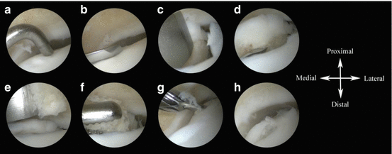
Fig. 8.2
Arthroscopic images of a left talus with a medial osteochondral defect that is treated by LDFF. (a) The exact location of the defect is identified by palpating the cartilage with a probe, while the ankle is in plantarflexion. (b) An osteochondral flap is created with the use of beaver knife. (c) With a chisel the osteochondral flap is lifted, taking care not to loosen the posterior part of the flap. (d) The bone flake of the osteochondral fragment is drilled with the use of a K-wire and a shaver blade to promote revascularization, again taking care not to loosen the fragment at its posterior attachment. (e) Cancellous bone is harvested from the distal tibia using a 4 mm chisel. (f) With an arthroscopic grasper the cancellous bone is transported to the defect until there is sufficient filling. (g) A cannulated system allows a predrilling and tapping of a compression screw (Arthrex Inc, Naples, USA). (h) An absorbable Bio-Compression screw (Arthrex Inc, Naples, USA) is placed 1–2 mm recessed relative to the surrounding surface of hyaline cartilage. The noncannulated screw is preferred because of its elegant diameter and compression strength
8.5 Postoperative Management
In both fixation techniques, a short-leg, non-weight-bearing cast will be applied for 4 weeks postoperatively. After these 4 weeks the foot is placed in a short-leg walking cast in neutral flexion position and neutral hindfoot position, with full weight bearing allowed. At 8 weeks postoperatively the cast will be removed. If a malleolar osteotomy was created during surgery, radiographs of the operated ankle are obtained to confirm consolidation. Physical therapy will be prescribed to assist in functional recovery and extend to full weight bearing in approximately 2 weeks.
8.6 Results
We published a retrospective case series of 9 patients after internal fixation of an OCD of the talus with a median follow-up of 4 years [10]. All patients had failed conservative treatment of a primary OCD. Various clinical outcome measures were recorded, including the Berndt and Harty outcome question [15]; Ogilvie-Harris score [16]; numeric rating scales (NRS; 0 to 10) of pain at rest, during walking and during running; American Orthopaedic Foot and Ankle Society (AOFAS; 0 to 100) score [17]; and Short Form 36 (SF-36; 0 to 100) [18, 19]. The Berndt and Harty clinical outcome was good in 7 cases (78 %) and fair in 2 cases (22 %). The Ogilvie-Harris score was excellent in 4 cases, good in 3 cases and fair in 2 cases. The median NRS pain at rest was 0 (range 0 to 6), during walking 1 (range 0 to 7) and during running 1.5 (range 0 to 4). The median AOFAS was 95 (range 77 to 100). The SF-36 physical component scale was 47.6 ± 8.3, and the mental component scale was 47.6 ± 13.9. On the final radiographs there were no progressive degenerative changes seen in all patients. An area of numbness around the scar was reported in one patient after fixation of the fragment.
Recently, we published the short-term clinical outcome of the arthroscopic LDFF technique for primary OCD of the talus, with a mean follow-up of 12 months (SD 0.6) [20]. Pre- and postoperative clinical assessment included the American Orthopaedic Foot and Ankle Society (AOFAS; 0 to 100) score [17] and the numeric rating scales (NRS; 0 to 100) of pain at rest and during walking. At final follow-up, the AOFAS score significantly improved from 63 ± 9.7 to 99 ± 1.6 (p < 0.001). The NRS of pain at rest significantly improved from 2.9 ± 1.9 to 0.1 ± 0.4 (p = 0.004) and during walking significantly improved from 7.6 ± 0.5 to 0.1 ± 0.4 (p < 0.001). On the final radiographs five of seven patients showed remodelling and bone ingrowth after LDFF (Fig. 8.3). All seven patients were satisfied and indicated that they would undergo the procedure again.
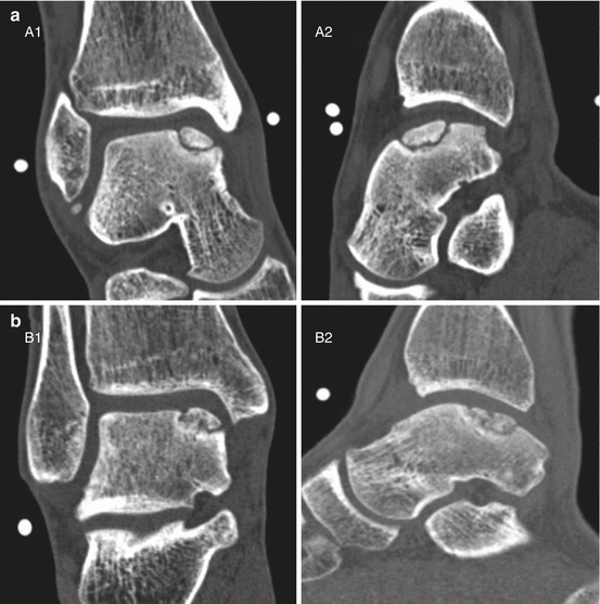
Fig. 8.3
 Rehabilitation and Return-to-Sports Activity After Debridement and Bone Marrow Stimulation of Osteochondral Talar Defects
Conservative Treatment of Talar Osteochondritis Dissecans (OCD)
Rehabilitation and Return-to-Sports Activity After Debridement and Bone Marrow Stimulation of Osteochondral Talar Defects
Conservative Treatment of Talar Osteochondritis Dissecans (OCD)
 Osteochondral Ankle Injuries in Footballers
 Management of Cystic Osteochondral Lesions of the Talus
 Mosaicplasty of Osteochondral Lesions of the Ankle
 Arthroscopic Debridement of Osteochondral Lesions of the Talus


(a) Preoperative coronal (A1) and sagittal (A2) computed tomography of a medial osteochondral talar defect of a right ankle in plantarflexion. (b) Postoperative coronal (B1) and sagittal (B2) computed tomography of the same ankle after LDFF with progressive bone ingrowth at 12-month follow-up
Related posts:
Rehabilitation and Return-to-Sports Activity After Debridement and Bone Marrow Stimulation of Osteochondral Talar Defects
Conservative Treatment of Talar Osteochondritis Dissecans (OCD)
Osteochondral Ankle Injuries in Footballers
Management of Cystic Osteochondral Lesions of the Talus
Mosaicplasty of Osteochondral Lesions of the Ankle
Arthroscopic Debridement of Osteochondral Lesions of the Talus
Stay updated, free articles. Join our Telegram channel
Full access? Get Clinical Tree
