, Paul D. Siney1 and Patricia A. Fleming1
(1)
The John Charnley Research Institute Wrightington Hospital, Wigan, Lancashire, UK
Leg length discrepancy, in general, and leg over-lengthening in particular, after total hip arthroplasty, has emerged as a topic of interest and dissatisfaction. In some quarters leg over-lengthening has been quoted as a common cause for litigation [1, 2]. “Limb lengthening is not uncommon after total hip replacement and may cause subjective problems for patients … 27 % of patients required heel lifts on the un-operated side” [3]. Leg length inequality “… as a cause of aseptic loosening, and unexplained pain, warrants investigation in THA patients” [4]. The high rate of dissatisfaction among patients with leg length inequality and the untoward results associated with this inequality, indicate that surgeons performing THA should familiarise themselves with a reliable method of equalising leg lengths intra-operatively” [5].
Ranawat and Rodriguez [6] attempted to assess functional leg length inequality (FLLI) by reviewing their records and gaining a response from the Hip Society members hoping to establish prevalence, aetiology and management. Fourteen percent of patients were noted to have pelvic obliquity and FLLI one month after surgery – all resolving by 6 months. Among the causes suggested by the respondents were: tightness of the periarticular tissues with resultant pelvic obliquity and degenerative conditions of the spine [6]. The direction of the pelvic obliquity, whether towards or away from the affected hip, was not stated. Reference to “tightness of the periarticular tissues” [6] and the fact that a proportion of patients needed a heel lift on the un-operated side [3] suggests that the tilt referred to was towards the symptomatic side thus highlighting abduction deformity of the osteoarthritic hip and functional limb lengthening on the symptomatic, arthritic side.
Despite the demand for THA the deformities associated with the arthritic hip have not received much attention. Lloyd-Roberts [7] in his Robert Jones Lecture suggested that fixed deformities in osteoarthritis of the hip are “due to capsular shortening reinforced by reflex guarding in the muscles supplied by the motor branches of the sensory nerves to the lower part of the cavity – such as pectineus, quadratus femoris and the adductor group”. This dynamic deformity was later followed by a structural deformity from the fibrosis of the musculo-tendinous function. Lloyd-Roberts felt that osteophytes, once developed, were unlikely to prevent movement by interlocking growth [7].
Pearson and Ridell [8] divided idiopathic osteoarthritic hips into two groups: the adducted and the non-adducted variety suggesting that muscle spasm was the primary cause, the final position depending on capsular contracture. (Mechanically these changes cannot develop in an ambulant patient, against gravity and deficient proximal lever – head and neck). Abduction deformity of a normal hip has been described with fibrous bands of genetic origin [9–11] or following intra-muscular injections [12]. The abducted position of the hip, usually with flexion and external rotation, is well recognised in acute poliomyelitis.
Why has the problem emerged after over 40 years of hip replacement surgery? Can the pitfalls be anticipated and, if not avoided, at least better understood and explained before surgery?
Patient Selection
Strict criteria of patient selection was the essential aspect in the evolution of the Charnley hip replacement. Significant pain at rest, failure of all conservative measures and an often radiologically destroyed hip, were the indications for hip arthroplasty. True limb shortening was, almost always, a part of the clinical picture. Radiologically upper pole grade III changes was the common finding [13]. The benefit of immediate pain relief far outweighed any possible disadvantage of failure to restore leg length. When this was achieved it was considered a bonus. So what has changed? Increasing experience with this type of surgery has led to an extension of the indications, increased the demand and higher patient expectations.
The term “end stage arthritis” has gained popularity but failed to define the clinical problem or the radiographic morphology of the osteoarthritic hip [13].
Surgical Technique
Limited exposures, excision of the capsule, laxity of the joint at trial reduction and the fear of post-operative dislocation all contribute to tight reduction – under anaesthetic. This is equated with stability – unfortunately at the expense of limb lengthening. Could this be avoided? Probably.
Excision of the Capsule
Excision of the capsule has never been a part of the Charnley technique. In fact preservation of the capsule was the teaching, and for very good reason. The hip capsule is not the site of the pain. Exposure, by trochanteric osteotomy, gives full access: a circumferential view of the acetabulum and access to the medullary canal. Checking stability at trial reduction, by joint distraction, offers a very definite endpoint. Excision of the capsule exposes the muscles which, under anaesthetic, allow a fair degree of stretch. This elastic “give” has no definite end-point, hence the need for lengthening, by component selection, and limb over-lengthening. Overstretching the muscles will lead to loss of proprioception, and even loss of power. Combined with the neuropathic nature of arthroplasty over-lengthening, laxity and muscle weakness would be opposite to the initial goal.
Component Selection
Pre-operative planning should include component selection as well as the detail of their positioning in relation to radiographic landmarks. With standard monoblock Charnley stem and the selection of mainly two cup sizes, the planning was focused on the details of the surgical technique. Modularity, although offering a wide choice, may result in the postponement of the decision until the final stage of the operation when both the cup and the stem are already securely fixed. The only remaining option will be the seating of the modular head on the neck taper. It is at this stage that leg length inequality, over-lengthening, may be the inevitable result.
Arthroplasty Technique
The advent of “minimally invasive” techniques and “resurfacing” must be considered. The techniques demand, ideally, anatomically intact, near normal geometry of the hip joint where limb shortening is not a problem, in fact in the early stages of arthritis, apparent, functional limb lengthening may be present. Maintaining limb length equality may be a problem and lengthening may result.
Patients at Risk for Limb Over-Lengthening
What has not received detailed attention is our ability to identify patients at risk for limb over-lengthening. Although anticipating the problem may not always be the solution, understanding the problem is the essential part of pre-operative planning and the informed consent.
Theoretical Considerations
With any painful condition it is natural to seek a position of comfort. In order to reduce the load on a painful hip, while standing, the pelvis is tilted to the symptomatic side, the hip is abducted, externally rotated, and the knee flexed. The contralateral hip, must of necessity, be adducted (observe Michelangelo’s David). While walking the body weight is moved over the symptomatic hip – reducing the load and the pain. These postural characteristics can only be adopted if certain mechanical features are present.
1.
Intact proximal lever. The arthritic hip must have the proximal lever, head and neck, relatively intact and contained with a normal or near normal acetabulum, allowing unhindered abduction movement. The radiographic appearances can be anticipated: incipient type arthritis, upper pole grade I and II, concentric, protrusio or medial pole type. Upper pole grade III type would be rare [13]. Inferior, medial osteophyte, “the head drop” is typical.
2.
The contralateral hip must allow adduction movement and thus be normal, near normal or be fixed in an adducted position.
3.
The spine must be mobile to compensate for the pelvic tilt. Alternatively, a fixed lateral spinal deformity, resulting in pelvic obliquity will be the primary cause of limb length inequality. The history of spinal problems should alert the surgeon to the possibility of functional limb length inequality, while the limb inequality and pelvic tilt should alert the surgeon to the likelihood of spinal problems.
4.
Soft tissue, capsular, contractures and osteophyte formation, must be secondary.
Clinical Relevance of Theoretical Considerations
With the information in mind it is possible to identify patients who are at risk for leg over-lengthening as a result of THA
Measurement of Limb Length Discrepancy
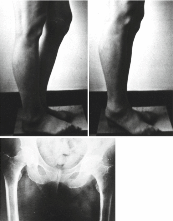
True and apparent leg length discrepancy (LLD) was measured by the standard, clinical methods. It was felt, however, that a functional leg length discrepancy (FLLD) would be of practical interest, because this is the discrepancy that affects patient’s gait and indicates the need and the extent of the footwear build-up (Figs. 5.1 and 5.2).


Figs. 5.1 and 5.2
Functional leg lengthening: abducted left hip, knee in flexion. With the knee extended right heel is off the ground. Radiograph shows pelvic tilt to the left side, well preserved proximal lever (head and neck) large infero-medial, “head drop” osteophyte – opposite hip normal
This measurement is taken with the patient erect. Theoretical considerations suggest that patients at risk for post-operative leg over-lengthening are those with pre-operative abduction deformity of the hip.
Stay updated, free articles. Join our Telegram channel

Full access? Get Clinical Tree


