Risk factors
Overuse in sport
Smoking
Obesity
Oral steroid use
Age 45–54 years
Other tendinopathies
Repetitive movement
Diabetes
White race
9.2 Anatomy
In order to better understand the etiology of this tendinopathy, it is essential to analyze the anatomic relationships of the lateral compartment of the elbow. Cohen and Romeo [9] showed the relationships that exist between the extensor carpi radialis longus (ECL) and ECRB. The ECRL origin is entirely muscular along the lateral supracondylar ridge of the humerus. The shape of the muscle is triangular, with the apex positioned proximally. Instead, the origin of the ECRB is entirely tendinous. Although the origin of the ECRB is mixed with that of the extensor digitorum communis, the authors showed [9] how dissecting from distal to proximal and following their under surface the two tendons can be isolated behind the humerus. The origin of the ECRB is located just below the distal-most tip of the lateral supracondylar ridge. The footprint of the tendon has a diamond shape of about 13 × 7 mm (Fig. 9.1). At the radiocapitellar joint, the tendon lies at the level of the front portion of the capsule, but it is possible to easily isolate the two structures at this level.
9.3 Pathomechanics
Biomechanical analysis has shown that eccentric contractions of the extensor carpi radialis brevis (ECRB) muscle during backhand tennis swings are the cause of repetitive microtraumas that result in microtears in the origin of the tendon [10]. Other authors have suggested different causes like direct trauma in the lateral region of the elbow or relative hypovascularity of the region, fluoroquinolone antibiotics, and anatomic predisposition [11–13]. Cyriax was the first to theorize that tears of the common extensor origin were involved in the disease process [14]. Subsequently, other authors showed that the nature of the disease is actually a degenerative tendinopathy. Goldie described the histological presence of granulation tissue found at the origin of the ECRB [15]. Macroscopic tearing in association with the histological findings was described by Coonrad and Hooper [6]. Nirschl called these histological changes “angiofibroblastic hyperplasia [16, 17].” In his study, he noted gray friable tissue characterized by disorganized collagen formation with immature fibroblastic and vascular elements. Subsequently, increased rates of apoptosis and cellular autophagy have been observed in tenocytes, resulting in disruption of extracellular collagen matrix and weakening of the tendon [18]. These changes at the tendon’s origin are the pathologic healing response to microtears caused by repetitive eccentric or concentric overloading of the extensor muscle mass [19]. Several studies have suggested that the origin of the extensor digitorum communis (EDC) is also implicated in lateral epicondylitis [20, 21].
9.4 Clinical Presentation
Patients complain of pain that radiates from the lateral epicondyle down to the forearm, often associated with weakness and difficulty in the handgrip.
Nirschl has divided symptoms into seven phases [22, 23]. A history of previous occurrence of tennis elbow also suggests tendinopathy. Imaging techniques such as magnetic resonance or diagnostic ultrasound are useful to identify the calcifications, tears, or ruptures of the ECRB [24, 25]. Physical examination should begin with cervical spine and be followed by the entire upper extremity. The examination proceeds then to the elbow. The elbow is tender over the lateral epicondyle and slightly distal, into the extensor mass.
Thomsen maneuver (resisted wrist extension with the elbow in full extension and forearm in pronation) or maximal wrist flexion can exacerbate pain at the lateral epicondyle. The first maneuver causes painful eccentric contraction at the origin of the ECRB. The second maneuver places the ECRB on maximal stretch, passively tensioning the muscle origin and thus causing pain. In order to exclude the presence of a plica, the elbow must be flexed passively with the forearm pronated and supinated. If a plica is involved, the point of maximal tenderness is usually located more distally and posteriorly, over the radiocapitellar joint, compared to lateral epicondylitis. Other causes of lateral sided elbow pain can be nerve entrapments at one or more sites, such as radial tunnel syndrome or posterior interosseous nerve (PIN) syndrome. Up to 5 % of patients with lateral epicondylitis presents radial nerve entrapment [26].
Pain elicited with resisted supination (when the nerve is trapped in the supinator muscle) or with resisted long-finger extension (when the nerve is trapped at the ECRB) can indicate PIN entrapment. Differential diagnosis between nerve entrapment and lateral epicondylitis can be difficult. The treatment of the two conditions is entirely different. The elbow examination is completed with a standard evaluation of elbow effusion, stability, and range of motion. The examination then moves distally toward the forearm and the hand. Grip strength should be tested to determine whether it decreases compared with the unaffected side or causes significant discomfort. Neurovascular status is a basic component of the examination and should be noted. Differential diagnosis for atraumatic lateral elbow pain may include radicular cervical spine disease, radial nerve compression, intra-articular loose bodies, and chondral lesions. Tumors, avascular necrosis, and osteochondritis dissecans of the capitellum, even if less common, may be considered as well (Table 9.2).
Table 9.2
Different diagnoses of lateral elbow pain
Pathology | History | Physical examination |
|---|---|---|
Cervical spondylosis | Neck pain Radicular pain to the elbow | Symptoms with spine compression extension |
Radial tunnel syndrome | Insidious pain at lateral elbow | Pain 2–4 cm distal to epicondyle |
Posterior interosseous nerve compression | Insidious pain at lateral elbow and weakness | Weakness of wrist and finger extensors |
Intra-articular bodies | Trauma | Clicking or limitation of range of motion |
Chondral lesions | Trauma | Clicking or limitation of range of motion |
Tumors | Night pain Prior malignancy | Palpable mass |
Avascular necrosis | Alcohol abuse HIV Sickle cell anemia Corticosteroids | Joint effusion, mechanical symptoms |
Osteochondritis dissecans | Gymnast Throwers, adolescent | Joint effusion, mechanical symptoms |
9.5 Surgical Treatment
Conservative treatment is the gold standard. However, between 5 % and 10 % of these patients develop persistent symptoms that may require surgical treatment. Particularly, persistent pain at night can determine the choice of surgical treatment. Surgical treatment with tendon release should be reserved in case of failure of the conservative treatment that should not last less than 6 months.
Surgical treatment can be percutaneous, open, or arthroscopic, with success rates ranging between 65 % and 95 % [27].
9.6 Percutaneous Treatment
A blade, often number 11, is inserted perpendicular to the skin anterior to the lateral epicondyle, then a one-centimeter-long skin incision is performed. A complete release of the common extensor origin is performed moving the tip of the blade anteriorly and inferiorly from the lateral epicondyle. A further displacement is then achieved by the Mill’s manipulation, consisting of a forcible, full extension of the elbow with the forearm fully pronated and the wrist and fingers held in flexion. At the end of the procedure, a gap of one centimeter, on average, is easily palpable between the lateral epicondyle and the retracted tendons. This procedure is preferred by many authors because it’s less invasive and the surgical results are similar to those of more elaborated procedures [28]. Baumgard and Schwartz [28] reported 91 % excellent (no symptoms under any circumstances), 0 % fair (improvement but still symptomatic), and 9 % unsatisfactory (no improvement) after an average follow-up of 34 months (range: 14–81 months). Another case series of percutaneous release reported similar results [29]. Powell and Burke [30] reviewed 20 patients after a follow-up raging from 5 to 36 months. They showed 85 % excellent or good results. Grundberg and Dobson [31] presented the results of percutaneous release in 32 cases of tennis elbow. With a mean follow-up of 26 months, he had 90.6 % of excellent and good results. More recently, Nazar et al. [32] showed how percutaneous release of the epicondylar muscles has a high rate of success: it is relatively simple to perform, it is done as a day-case procedure, and it doesn’t show complications. As a matter of fact, the postoperative outcome was between good and excellent in most patients. Eighty-seven percent of patients had complete pain relief with no complications reported.
All the patients returned to their normal jobs and hobbies such as gardening, horse riding, and playing musical instruments. We personally do not routinely choose this technique because of the theoretical risk of damaging part of the radial component of the lateral collateral ligament, although this is not supported by the available literature.
9.7 Open Treatment
Several open techniques have been described. The original technique (1955 Bosworth) [33] involves the identification and removal of the abnormal tissue that surrounds the common origin of the extensor tendons, the creation of a bone bed that promotes healing, and finally the reconstruction of the overlying aponeurosis. First of all, it is necessary to identify the ERCB tendon: its origin is located below the lateral epicondylar prominence, along a longitudinal ridge, and is directed from the upper part of the capitellum to the level of the radiohumeral joint.
Its tendon runs below the extensor digitorum communis and its aponeurosis, distally to the epicondyle. It can be easily isolated, proceeding from anterior to posterior and starting at the junction between the ECRL and EDC aponeurosis. The undersurface of the ECRB tendon can be elevated from the ECRL muscle in oblique fashion. The aponeurosis of the EDC lies on top of the ECRB and is tightly opposed. The ECRB tendon is debrided and the epicondylar origin denuded or drilled. The open approach leads to greater visualization of the operative field and the pathologic tissue; however, it is associated with a higher incidence of complications and a longer time to return to work [34].
9.8 Arthroscopic Treatment
Arthroscopic release is especially indicated when a concomitant intra-articular pathology is suspected. The advantage of exploring the joint is recently increasing the indications. Patient is placed in lateral decubitus position with the operative arm supported by an arm holder at 100° of flexion/90° of abduction at the level of the shoulder. The elbow is positioned at 90° of flexion, with the forearm hanging free from gravity (Fig. 9.2). Forty milliliter of sterile saline solution is injected before placing the portals to distend the elbow joint. A proximal anteromedial portal is created 2–3 cm proximal to the medial humeral epicondyle and 1 cm anterior to the intramuscular septum. Then a 30° arthroscope is inserted into this portal. This allows intra-articular diagnostic evaluation of the anterior compartment. The proximal anterolateral portal is located approximately 3 cm proximal and 1 cm anterior to the lateral epicondyle. A retractor, aimed at the radiocapitellar joint to protect the posterior interosseous nerve, can be inserted through this portal. The instruments are introduced through the anterolateral portal, located 1.5–2 cm proximal and 1 cm anterior to the lateral epicondyle.
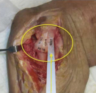
Fig. 9.1


Cadaveric preparation in right elbow: in the yellow circle the anatomy of LCL complex; the yellow arrow shows ECRB and close relation with R-LCL (An Lig. = Annular ligament)
Related posts:


Stay updated, free articles. Join our Telegram channel
Full access? Get Clinical Tree
