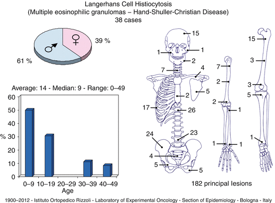
Location: Flat and short bones of the trunk: skull (parietal, frontal site), ribs, pelvis, vertebral body, clavicle, and scapula. Among long bones: proximal half of the femur, humerus, and tibia (diaphyseal localization is typical). Very rare in the hand and foot.
Clinical: Pain, wide swelling in superficial bones, rare pathologic fracture, radicular pain, rarely signs of medullary compression, and deformities of the column in vertebral lesions. Rare mild increase in sedimentation rate and mild eosinophilia.
Imaging: Standard X-rays show an osteolytic lesion with variable features. Sometimes rounded, often polycyclic, well-defined margins, thin sclerotic rim, and overall benign-looking appearance. Sometimes “moth-eaten” pattern with ill-defined margins, no sclerotic rim, and “onionskin” periosteal reaction mimicking a malignant process (e.g., Ewing sarcoma). Uniform, rapid flattening of the vertebral body reduced to a thin bony lamina (vertebra plana) is typical. Bone scan sometimes shows multiple lesions. On MRI light gray, intermediate low signal on T1, and a higher than fat, bright signal on T2.
Pathology: Soft, semiliquid, yellowish gray, with areas of hemorrhage or necrosis. Background of large pale-staining cells (histiocytes) with infiltration of leukocytes, without intercellular matrix, punctuated by nodules of small eosinophilic cells. Histiocytes have wide cytoplasm, with ill-defined membrane, with reniform, indented, pale nucleus, small nucleolus, and may be collected in nests or nodules or form a more rarefied background to the infiltration of eosinophiles with small lobate, dark nucleus, and cytoplasm stuffed with bright red granules. Few neutrophiles and lymphocytes. Abundant reticulum surrounding small group of cells. Sporadic giant cells, foam cells, mitotic figures.
Course and Staging: Rapid growth, spontaneously self-limiting and tendency to healing with at least partial bone repair, rare evolution into multiple type, exceptional transformation into chronic diffused type. Usually, stage 2, rarely stage 3.
Treatment: Needle biopsy, frozen section, and steroid injection treatment of choice. Clinically very successful with complete or almost complete repair in 2 years. Bracing or casting required in the spine. Radiotherapy may be used (2–3,000 r). Systemic cortisone and chemotherapy are used in multiple lesions.


Key Points
Clinical | Pain, swelling, rapidly increasing
Stay updated, free articles. Join our Telegram channel
Full access? Get Clinical Tree
 Get Clinical Tree app for offline access

|


