Chapter 7 Knee and Thigh
The knee is a modified, hinged weight-bearing joint dependent on several structures for stability:
• medial and lateral collateral ligaments to prevent lateral (sideways) shearing movements
• anterior and posterior cruciate ligaments to prevent anterior (forward) and posterior (backwards) displacement during movement
• menisci (two wedge-shaped cartilages) form mechanical spacers to cushion forces, guide movements and add to overall stability.
• heavy weight-bearing demands on the knee during sporting activities
• mechanical disadvantages at extremes of performance, e.g. a relatively weak medial collateral ligament and its attachment to the medial meniscus
• extremes of force as seen in tackling that can lead to severe injuries such as anterior cruciate ligament ruptures.
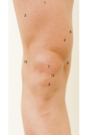
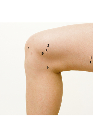
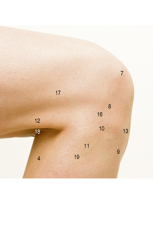
Behind the knee on the lateral side, the rounded tendon of biceps femoris (12) can be felt easily, with the broad, strap-like iliotibial tract (17) in front of it, with a furrow between them. On the medial side, two tendons can be felt: the narrow rounded semitendinosus (14) just behind the broader semimembranosus (5). At the front, the patellar ligament (13) keeps the patella (7) at a constant distance from the tibial tuberosity (9), while at the side the adjacent margin of the femoral condyle and tibial plateau can be palpated.
ANATOMICAL AREA: KNEE AND THIGH
TAPING FOR MEDIAL COLLATERAL LIGAMENT KNEE SPRAIN
Indications for use
• MCL sprains: 1st and 2nd degree
• post immobilization of 3rd-degree MCL sprains
• for medial meniscus injuries: emphasize spiral strips which cause internal rotation of the tibia
• Ensure that the correct diagnosis has been made. If in doubt, refer!
• Always assess which knee and which side of that knee was injured (the athlete may have sustained spraining on the medial aspect and bruising on the lateral aspect of the same knee).
• During exercise, increased blood circulation causes swelling of thigh muscles. Ensure that circumferential strips are affixed with only light tension, avoiding constriction and possible cramping of thigh and calf muscles.
• Avoid taping over the patella, as this can cause compression, pain and subsequent problems.
For additional details regarding an injury example see T.E.S.T.S. chart, p. 144.
Positioning
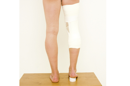
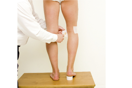
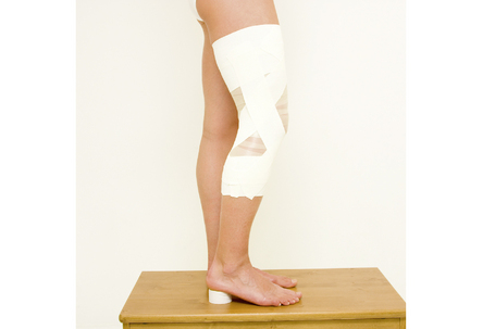
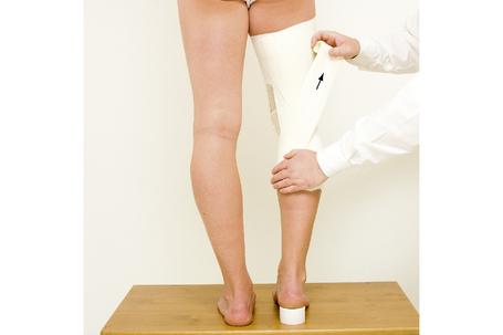
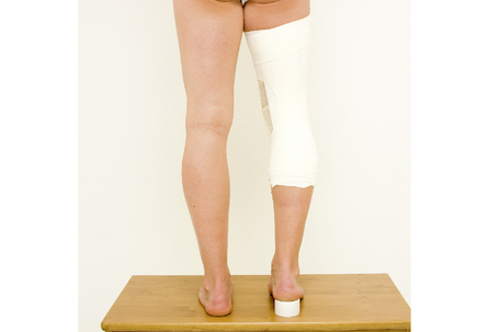
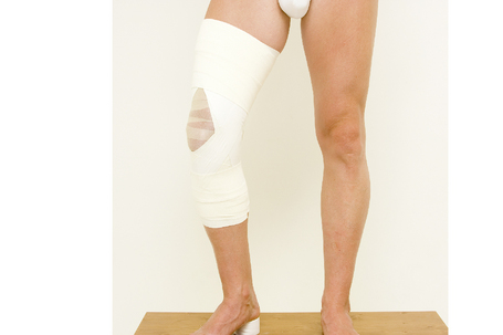
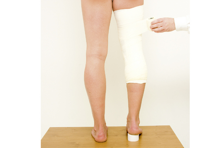
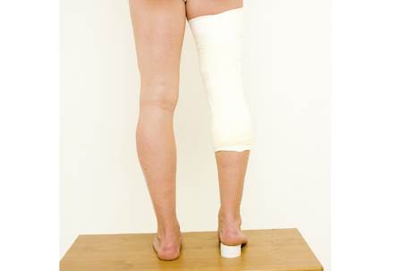
Standing upright. Place a roll of tape under the heel of the foot of the injured knee, so that the knee is slightly flexed. The foot is turned inwards to medially rotate the tibia under the femur (releasing the tension from the MCL). Eighty percent of the body weight should be supported by the uninjured side.
Procedure
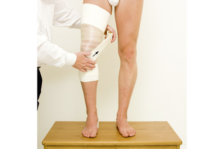
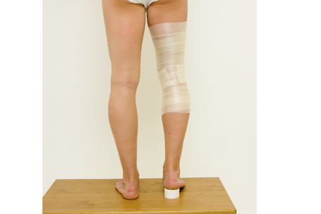
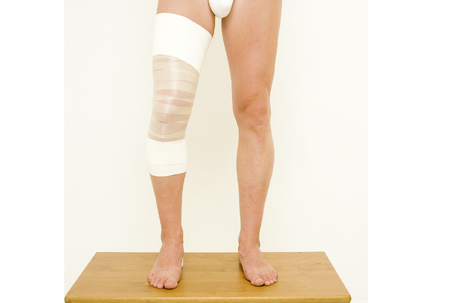
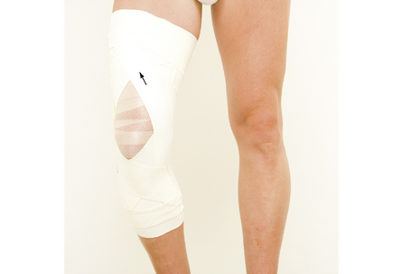
7. Place a strip of 7.5 cm elastic adhesive bandage starting from the posteromedial aspect of the distal anchor, spiralling around the lateral side of the tibia, anteriorly. Cross medially below the patella with moderate tension and pull proximally with strong tension over the medial joint line to the proximal anchor.
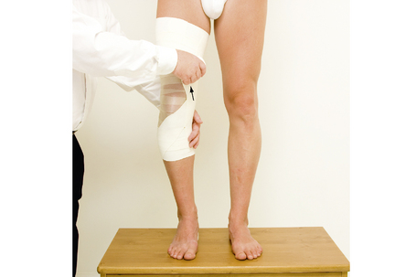
8. Place the second strip of 7.5 cm elastic adhesive bandage also starting at the posteromedial aspect of the distal anchor, this time heading anteriorly on the medial side. Pull proximately with strong tension over the medial joint line to the proximal anchor anteriorly, releasing the tension only when adhering the end of the strip.
ANATOMICAL AREA: KNEE AND THIGH
INJURY: KNEE SPRAIN MEDIAL COLLATERAL LIGAMENT
• medial collateral ligament sprain: 1st–3rd degree of severity (see Sprains chart, p. 36)
• internal collateral ligament sprain: 1st–3rd degree of severity
• excessive inward pressure forcing the knee medially into valgus (inwardly bent ‘knock-kneed’ position). Example: a football player tackled at the knees from the left side may sustain a medial sprain of the left knee and potentially a lateral sprain of the right knee
• sudden impact forcing body laterally on a fixed foot
• often associated with other injured structures (medial meniscus, medial collateral ligament and anterior cruciate ligament: ‘the unhappy triad’)
• local pain and tenderness on the medial side (inside) of the knee
• active movement testing: medial pain on end-range extension
• resistance testing (neutral position): no pain on moderate resistance
a. 1st- and 2nd-degree sprain: medial pain with or without instability when tested at 30 ° knee flexion
b. 3rd degree of severity: complete ligament rupture ‘opens up’ at 30 °, can be less pain than with 2nd degree
c. instability at 0 ° extension is indicative of a severe injury with posterior capsule involvement
• continued therapy including:
< div class='tao-gold-member'>
Stay updated, free articles. Join our Telegram channel

Full access? Get Clinical Tree


 NOTES:
 TIP:
TIP: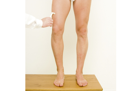
 NOTE:
NOTE: NOTE:
NOTE: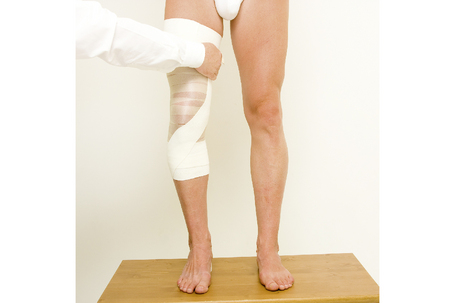
 TIP:
TIP: NOTE:
NOTE: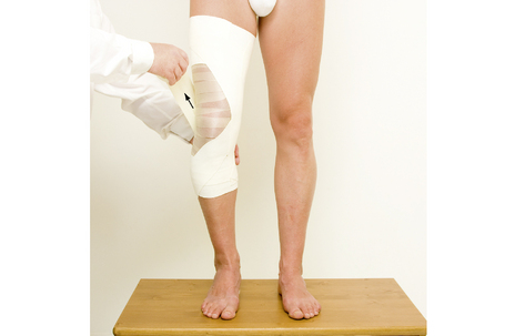
 NOTE:
NOTE: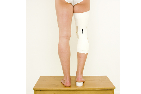
 NOTE:
NOTE: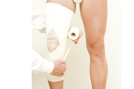
 TIP:
TIP: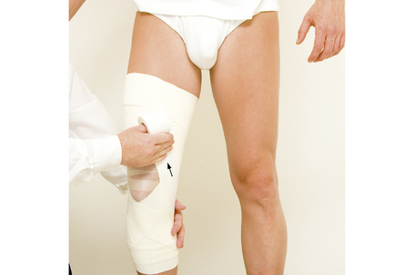
 TIP:
TIP: NOTE:
NOTE: NOTE:
NOTE: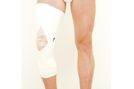
 NOTE:
NOTE: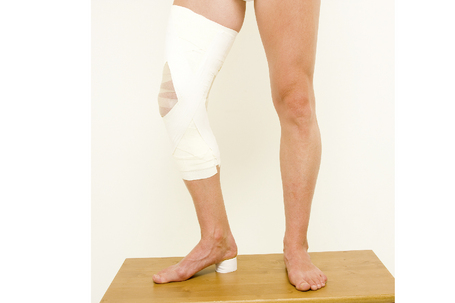
 NOTES:
 NOTE:
NOTE:


